File:Keibel Mall 2 032.jpg
From Embryology

Size of this preview: 800 × 500 pixels. Other resolution: 1,200 × 750 pixels.
Original file (1,200 × 750 pixels, file size: 139 KB, MIME type: image/jpeg)
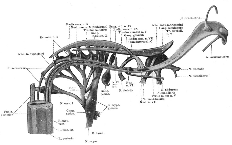
Fig. 31. Reconstruction showing the cranial nerves in a 10 mm human embryo
(Huber collection No. 3).
The brain wall is removed so as to show the primary sensory paths and motor nuclei of the different nerves.
File history
Yi efo/eka'e gwa ebo wo le nyangagi wuncin ye kamina wunga tinya nan
| Gwalagizhi | Nyangagi | Dimensions | User | Comment | |
|---|---|---|---|---|---|
| current | 12:57, 24 Ocak 2014 | | 1,200 × 750 (139 KB) | Z8600021 (talk | contribs) | ==Fig. 31. Reconstruction showing the cranial nerves in a 10 mm human embryo== (Huber collection No. 3). The brain wall is removed so as to show the primary sensory paths and motor nuclei of the different nerves. |
You cannot overwrite this file.
File usage
The following 3 pages use this file: