File:Placenta lobule blood flow cartoon.jpg

Original file (692 × 800 pixels, file size: 134 KB, MIME type: image/jpeg)
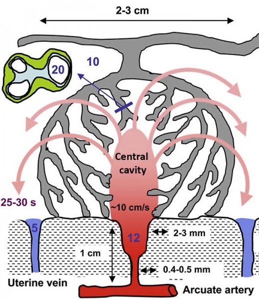
Placenta Lobule Blood Flow
Values were originally predicted in the reference paper by modelling and diagram not to scale. Dilation of the distal segment in normal pregnancies will reduce the velocity of incoming blood, and the residual momentum will carry the blood into the central cavity of the lobule, from where it will disperse evenly through the villous tree.
Transit time to the uterine vein is estimated to be in the order of 25–30 s, allowing adequate time for oxygen exchange.
The pressure of the maternal blood, indicated in mmHg by the figures in blue, will drop across the non-dilated segment of the spiral artery, the dimensions of which are given alongside.
- Links: Placenta lobule blood flow cartoon | Figure - Uterine and placental vasculature | Figure - Placenta spiral artery conversion | Placenta Development
Reference
<pubmed>19375795</pubmed>| PMC2697319
Placenta. 2009 June; 30(6): 473–482. doi: 10.1016/j.placenta.2009.02.009.
Copyright
© 2009 Elsevier Ltd. “This is an unofficial translation of an article that appeared in an Elsevier publication. Elsevier has not endorsed this translation.”
http://www.elsevier.com/wps/find/authorsview.authors/supplementalterms1.0
Original figure has been, cropped, relabelled and resized.
File history
Click on a date/time to view the file as it appeared at that time.
| Date/Time | Thumbnail | Dimensions | User | Comment | |
|---|---|---|---|---|---|
| current | 19:27, 6 November 2013 | | 692 × 800 (134 KB) | Z8600021 (talk | contribs) | ==Placenta Lobule Blood Flow== Values were originally predicted in the paper by modelling and diagram not to scale. Dilation of the distal segment in normal pregnancies will reduce the velocity of incoming blood, and the residual momentum will carry t... |
You cannot overwrite this file.
File usage
There are no pages that use this file.