File:DeBruin1910 fig10.jpg

Original file (1,422 × 1,100 pixels, file size: 208 KB, MIME type: image/jpeg)
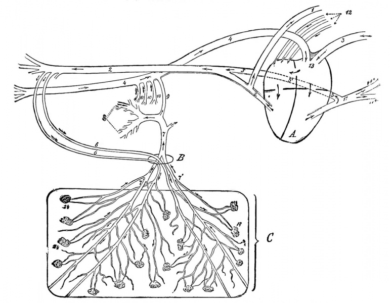
Fig. 10. Schematic Sketch of the Foetal Circulation of a Calf.
The arrows indicate the direction in which the blood flows.
A, Heart; B, umbilical opening; C, portion of the chorion. 1, Anterior aorta; 2, posterior aorta; 3 anterior vena cava; 4, posterior vena cava; 5, duct of Botalli; part of Botalli's duct posterior to the heart (sketched somewhat too long, but was necessary in order to demonstrate it) ; 6, umbilical arteries; 7, umbilical vein; 7', some of its branches; 8, portal vein; 9, ductus venosus; 10, portal veins: 11, pulmonary artery; 11', some of its branches; 12, pulmonary veins; 13, tuberculum Loweri; 14, chorion papillae.
- Links: Bovine Development | Placenta Development
| Historic Disclaimer - information about historic embryology pages |
|---|
| Pages where the terms "Historic" (textbooks, papers, people, recommendations) appear on this site, and sections within pages where this disclaimer appears, indicate that the content and scientific understanding are specific to the time of publication. This means that while some scientific descriptions are still accurate, the terminology and interpretation of the developmental mechanisms reflect the understanding at the time of original publication and those of the preceding periods, these terms, interpretations and recommendations may not reflect our current scientific understanding. (More? Embryology History | Historic Embryology Papers) |
File history
Yi efo/eka'e gwa ebo wo le nyangagi wuncin ye kamina wunga tinya nan
| Gwalagizhi | Nyangagi | Dimensions | User | Comment | |
|---|---|---|---|---|---|
| current | 03:49, 4 November 2013 |  | 1,422 × 1,100 (208 KB) | Z8600021 (talk | contribs) | ==Fig. 10== {{Historic Disclaimer}} |
You cannot overwrite this file.
File usage
The following page uses this file:
