File:Hydatidiform mole 01.jpg
From Embryology
No higher resolution available.
Hydatidiform_mole_01.jpg (800 × 592 pixels, file size: 634 KB, MIME type: image/jpeg)
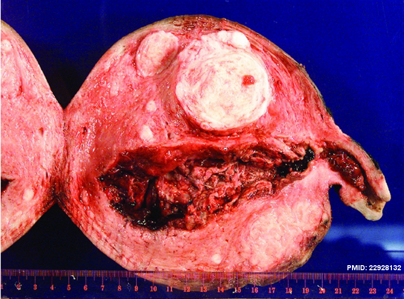
Figure 2: Pathology specimen of entire gross uterus and molar component within the lower uterine segment extending through the internal cervical os. Normal placental bed visualized within the fundal portion of the endometrial cavity. Multiple fibroids seen within the uterus.
File history
Yi efo/eka'e gwa ebo wo le nyangagi wuncin ye kamina wunga tinya nan
| Gwalagizhi | Nyangagi | Dimensions | User | Comment | |
|---|---|---|---|---|---|
| current | 22:14, 10 Haziran 2013 | | 800 × 592 (634 KB) | Z8600021 (talk | contribs) | Figure 2: Pathology specimen of entire gross uterus and molar component within the lower uterine segment extending through the internal cervical os. Normal placental bed visualized within the fundal portion of the endometrial cavity. Multiple fibroids ... |
You cannot overwrite this file.
File usage
The following page uses this file:


