File:Placenta previa and increta 02.jpg
Placenta_previa_and_increta_02.jpg (800 × 544 pixels, file size: 48 KB, MIME type: image/jpeg)
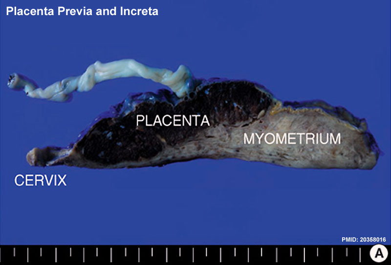
Placenta previa and Increta
Cut surface of the uterus with attached placenta and umbilical cord.
The left end of the uterus is the uterine cervix, and the right end of uterus is the uterine fundus. The cut surface shows abnormal placental adherence in the low uterine segment (placenta previa). The placenta invades into the myometrium, but does not penetrate through it (placenta increta).
Reference
<pubmed>20358016</pubmed>| PMC2844598 | J Korean Med Sci.
Copyright
This is an Open Access article distributed under the terms of the Creative Commons Attribution Non-Commercial License (http://creativecommons.org/licenses/by-nc/3.0) which permits unrestricted non-commercial use, distribution, and reproduction in any medium, provided the original work is properly cited.
Fig. 3 Jkms-25-651-g003-l.jpg http://synapse.koreamed.org/ViewImage.php?Type=F&aid=263059&id=F3&afn=63_JKMS_25_4_651&fn=jkms-25-651-g003_0063JKMS
Panel A cropped and resized from original figure.
File history
Yi efo/eka'e gwa ebo wo le nyangagi wuncin ye kamina wunga tinya nan
| Gwalagizhi | Nyangagi | Dimensions | User | Comment | |
|---|---|---|---|---|---|
| current | 11:17, 4 Haziran 2012 | | 800 × 544 (48 KB) | Z8600021 (talk | contribs) | ==Placenta previa and Increta== Cut surface of the uterus with attached placenta and umbilical cord. The left end of the uterus is the uterine cervix, and the right end of uterus is the uterine fundus. The cut surface shows abnormal placental adherence |
You cannot overwrite this file.


