File:Rugh 132.jpg
From Embryology
Size of this preview: 441 × 600 pixels. Other resolution: 735 × 1,000 pixels.
Original file (735 × 1,000 pixels, file size: 195 KB, MIME type: image/jpeg)
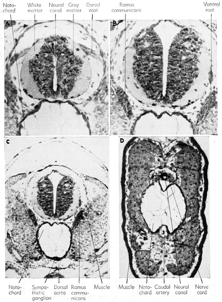
Development of the spinal cord and sympathetic nerves
(A) Showing the dorsal root (from neural crest).
(B) Showing the ventral root (from cord neuroblasts).
(C) Showing connection between the spinal nerve and the sympa- thetic ganglion, known as the ramus communicans.
(D) A section in the tail region to show the relatively large notochord and reduced nerve cord.
File history
Yi efo/eka'e gwa ebo wo le nyangagi wuncin ye kamina wunga tinya nan
| Gwalagizhi | Nyangagi | Dimensions | User | Comment | |
|---|---|---|---|---|---|
| current | 18:59, 14 April 2013 | | 735 × 1,000 (195 KB) | Z8600021 (talk | contribs) |
You cannot overwrite this file.
File usage
The following 2 pages use this file:


