File:Gastrointestinal villi and crypts cartoon.jpg
From Embryology
Gastrointestinal_villi_and_crypts_cartoon.jpg (500 × 333 pixels, file size: 28 KB, MIME type: image/jpeg)
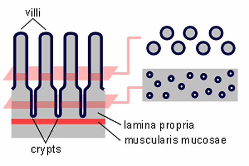
Gastrointestinal villi and crypts cartoon
The entire intestinal mucosa forms intestinal villi (about one mm long), which increase the surface area by a factor of ~ ten. The surface of the villi is formed by a simple columnar epithelium. Each absorptive cell or enterocyte of the epithelium forms numerous microvilli (1 µm long and about 0.1 µm wide). Microvilli increase the surface area by a factor of ~ 20.
File history
Click on a date/time to view the file as it appeared at that time.
| Date/Time | Thumbnail | Dimensions | User | Comment | |
|---|---|---|---|---|---|
| current | 13:48, 18 February 2013 |  | 500 × 333 (28 KB) | Z8600021 (talk | contribs) | ==Gastrointestinal villi and crypts cartoon== The entire intestinal mucosa forms intestinal villi (about one mm long), which increase the surface area by a factor of ~ ten. The surface of the villi is formed by a simple columnar epithelium. Each absorpti |
You cannot overwrite this file.
File usage
The following page uses this file:
