File:Gray0608.jpg
Original file (400 × 623 pixels, file size: 74 KB, MIME type: image/jpeg)
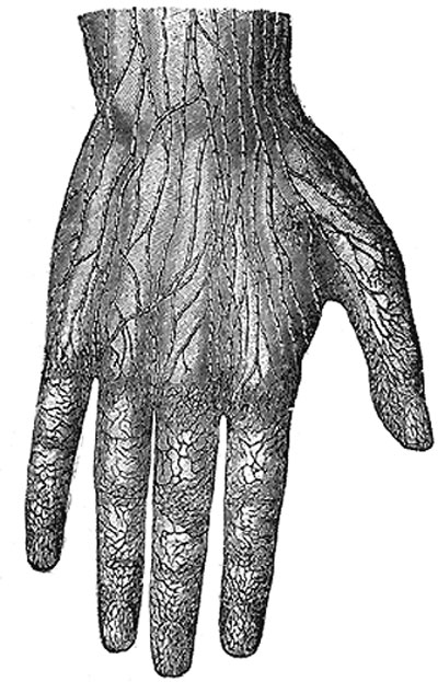
Lymphatic vessels of the dorsal surface of the hand
(Sappey.)
The superficial lymphatic vessels commence (Fig. 608) in the lymphatic plexus which everywhere pervades the skin; the meshes of the plexus are much finer in the palm and on the flexor aspect of the digits than elsewhere. The digital plexuses are drained by a pair of vessels which run on the sides of each digit, and incline backward to reach the dorsum of the hand. From the dense plexus of the palm, vessels pass in different directions, viz., upward toward the wrist, downward to join the digital vessels, medialward to join the vessels on the ulnar border of the hand, and lateralward to those on the thumb. Several vessels from the central part of the plexus unite to form a trunk, which passes around the metacarpal bone of the index finger to join the vessels on the back of that digit and on the back of the thumb. Running upward in front of and behind the wrist, the lymphatic vessels are collected into radial, median, and ulnar groups, which accompany respectively the cephalic, median, and basilic veins in the forearm. A few of the ulnar lymphatics end in the supratrochlear glands, but the majority pass directly to the lateral group of axillary glands. Some of the radial vessels are collected into a trunk which ascends with the cephalic vein to the deltoideopectoral glands; the efferents from this group pass either to the subclavicular axillary glands or to the inferior cervical glands.
- Gray's Images: Development | Lymphatic | Neural | Vision | Hearing | Somatosensory | Integumentary | Respiratory | Gastrointestinal | Urogenital | Endocrine | Surface Anatomy | iBook | Historic Disclaimer
| Historic Disclaimer - information about historic embryology pages |
|---|
 |
| iBook - Gray's Embryology | |
|---|---|

|
|
Reference
Gray H. Anatomy of the human body. (1918) Philadelphia: Lea & Febiger.
Cite this page: Hill, M.A. (2024, June 16) Embryology Gray0608.jpg. Retrieved from https://embryology.med.unsw.edu.au/embryology/index.php/File:Gray0608.jpg
- © Dr Mark Hill 2024, UNSW Embryology ISBN: 978 0 7334 2609 4 - UNSW CRICOS Provider Code No. 00098G
File history
Click on a date/time to view the file as it appeared at that time.
| Date/Time | Thumbnail | Dimensions | User | Comment | |
|---|---|---|---|---|---|
| current | 01:10, 15 February 2013 | | 400 × 623 (74 KB) | Z8600021 (talk | contribs) | ==Lymphatic vessels of the dorsal surface of the hand== (Sappey.) |
You cannot overwrite this file.
File usage
The following 3 pages use this file: