File:Gray0606.jpg
Original file (686 × 800 pixels, file size: 93 KB, MIME type: image/jpeg)
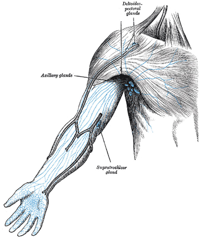
The Lymph Glands of the Upper Extremity
The superficial lymph glands and lymphatic vessels of the upper extremity.
The lymph glands of the upper extremity are divided into two sets, superficial and deep.
The superficial lymph glands are few and of small size. One or two supratrochlear glands are placed above the medial epicondyle of the humerus, medial to the basilic vein. Their afferents drain the middle, ring, and little fingers, the medial portion of the hand, and the superficial area over the ulnar side of the forearm; these vessels are, however, in free communication with the other lymphatic vessels of the forearm. Their efferents accompany the basilic vein and join the deeper vessels. One or two deltoideopectoral glands are found beside the cephalic vein, between the Pectoralis major and Deltoideus, immediately below the clavicle. They are situated in the course of the external collecting trunks of the arm.
(Text from Gray's Anatomy 1918)
- Gray's Images: Development | Lymphatic | Neural | Vision | Hearing | Somatosensory | Integumentary | Respiratory | Gastrointestinal | Urogenital | Endocrine | Surface Anatomy | iBook | Historic Disclaimer
| Historic Disclaimer - information about historic embryology pages |
|---|
 |
| iBook - Gray's Embryology | |
|---|---|

|
|
Reference
Gray H. Anatomy of the human body. (1918) Philadelphia: Lea & Febiger.
Cite this page: Hill, M.A. (2024, June 21) Embryology Gray0606.jpg. Retrieved from https://embryology.med.unsw.edu.au/embryology/index.php/File:Gray0606.jpg
- © Dr Mark Hill 2024, UNSW Embryology ISBN: 978 0 7334 2609 4 - UNSW CRICOS Provider Code No. 00098G
File history
Yi efo/eka'e gwa ebo wo le nyangagi wuncin ye kamina wunga tinya nan
| Gwalagizhi | Nyangagi | Dimensions | User | Comment | |
|---|---|---|---|---|---|
| current | 16:11, 14 February 2013 | | 686 × 800 (93 KB) | Z8600021 (talk | contribs) | ==The Lymph Glands of the Upper Extremity== The superficial lymph glands and lymphatic vessels of the upper extremity. The Lymph Glands of the Upper Extremity (Fig. 606).—The lymph glands of the upper extremity are divided into two sets, superficial a |
You cannot overwrite this file.
File usage
The following 3 pages use this file: