File:Tetralogy of Fallot 01.jpg
Original file (700 × 873 pixels, file size: 113 KB, MIME type: image/jpeg)
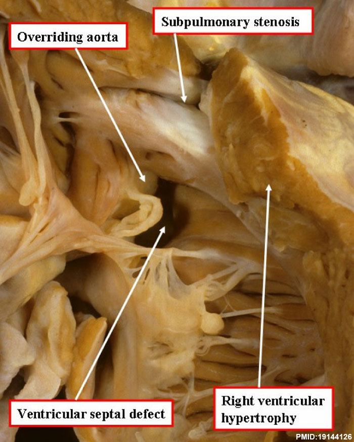
Tetralogy of Fallot
An autopsied specimen has been opened through the anterior wall of the right ventricle to show the cardinal features of tetralogy of Fallot.
Reference
Bailliard and Anderson Orphanet Journal of Rare Diseases 2009 4:2 doi:10.1186/1750-1172-4-2
Copyright
© 2009 Bailliard and Anderson; licensee BioMed Central Ltd. This is an Open Access article distributed under the terms of the Creative Commons Attribution License (http://creativecommons.org/licenses/by/2.0), which permits unrestricted use, distribution, and reproduction in any medium, provided the original work is properly cited.
1750-1172-4-2-1.jpg
File history
Yi efo/eka'e gwa ebo wo le nyangagi wuncin ye kamina wunga tinya nan
| Gwalagizhi | Nyangagi | Dimensions | User | Comment | |
|---|---|---|---|---|---|
| current | 23:47, 22 January 2013 | | 700 × 873 (113 KB) | Z8600021 (talk | contribs) | ==Tetralogy of Fallot== An autopsied specimen has been opened through the anterior wall of the right ventricle to show the cardinal features of tetralogy of Fallot. ===Reference=== Bailliard and Anderson Orphanet Journal of Rare Diseases 2009 4:2 doi: |
You cannot overwrite this file.
File usage
The following page uses this file: