File:Wislocki1920 plate 4.jpg

Original file (935 × 1,000 pixels, file size: 199 KB, MIME type: image/jpeg)
Plate 4
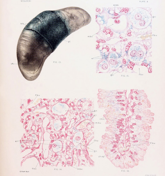
Fig. 13. Cat fetus, measuring 6.8 cm., surrounded by unruptured membranes, showing the coloration of the placenta and chorion after repeated injection of trypan-blue into the maternal circulation. Note that the chorion over the poles of the fetus is unstained. The allantoic and amniotic fluids do not contain a trace of dye and the fetus is unstained.
Fig. 14. Placenta of cat, nearly full term, after repeated injection of trypan-blue into the maternal blood stream, showing the distribution of dye in the chorionic epithelium.
Fig. 15. Placenta of a vitally stained cat, nearly full term, showing several multimiclear giant cells of the chorionic ectoderm filled with particles of trypan-blue.
Fig. 16. Section of the "brown border" of the placenta of a cat, nearly full term, showing the absorption of erythrocytes and of trypan-blue by the chorionic epithelium.
File history
Yi efo/eka'e gwa ebo wo le nyangagi wuncin ye kamina wunga tinya nan
| Gwalagizhi | Nyangagi | Dimensions | User | Comment | |
|---|---|---|---|---|---|
| current | 17:45, 27 December 2012 |  | 935 × 1,000 (199 KB) | Z8600021 (talk | contribs) |
You cannot overwrite this file.
File usage
The following 2 pages use this file: