File list
From Embryology
This special page shows all uploaded files.
| Date | Name | Thumbnail | Size | User | Description | Versions |
|---|---|---|---|---|---|---|
| 20:45, 2 March 2020 | Beattie1924 fig02.jpg (file) |  |
194 KB | Z8600021 | 2 | |
| 20:44, 2 March 2020 | Beattie1924 fig01.jpg (file) |  |
238 KB | Z8600021 | 2 | |
| 12:12, 28 February 2020 | 1897 Morgan - The development of the frog's egg.pdf (file) |  |
15.5 MB | Z8600021 | ==The Development of the Frog's Egg== '''An Introduction to Experimental Embryology''' by Thomas Hunt Morgan {{Morgan1897 footer}} Category:PDF | 1 |
| 12:11, 28 February 2020 | 1913 Morgan - Heredity and sex.pdf (file) | |
17.9 MB | Z8600021 | ==Heredity and Sex (1913)== Category:PDF | 1 |
| 18:42, 26 February 2020 | Max Cat.jpg (file) |  |
180 KB | Z8600021 | 1 | |
| 18:39, 26 February 2020 | Max cat.jpg (file) |  |
104 KB | Z8600021 | 1 | |
| 15:09, 25 February 2020 | Hypothalamus model 01.jpg (file) |  |
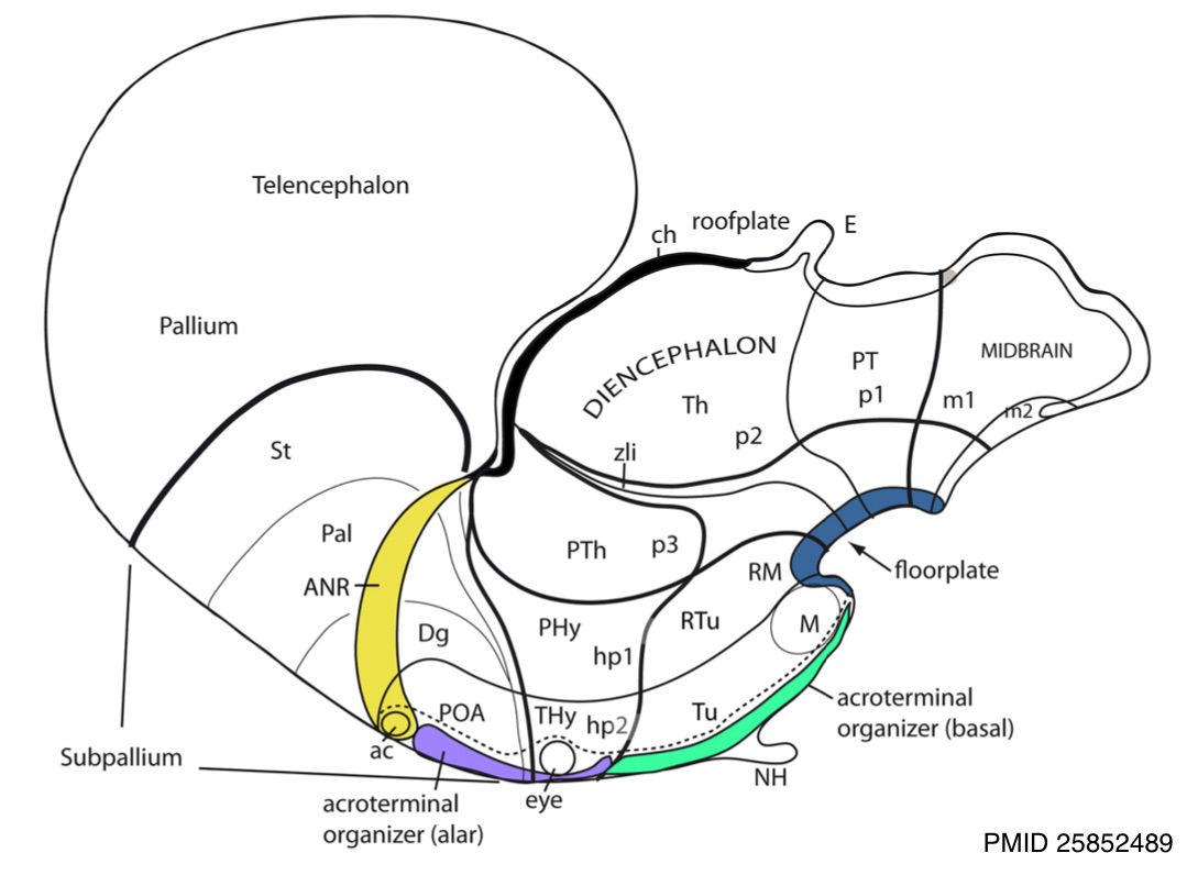
119 KB | Z8600021 | ==Hypothalamus Model== Apparent sources of patterning diffusible morphogens that may have effects on the hypothalamus. Anterior neural ridge (ANR; yellow), which releases {{FGF}}8 is in fact a part of the roof plate (dorsalizing influence), rather than a source of AP effects. in contrast, the retromamillary and mamillary floor plate (dark blue associated to RM and M) releases {{SHH}} (ventralizing influence; note Shh secondarily also is expressed throughout the basal plate, and is later d... | 1 |
| 14:05, 25 February 2020 | Brain stem subdivisions 01.jpg (file) |  |
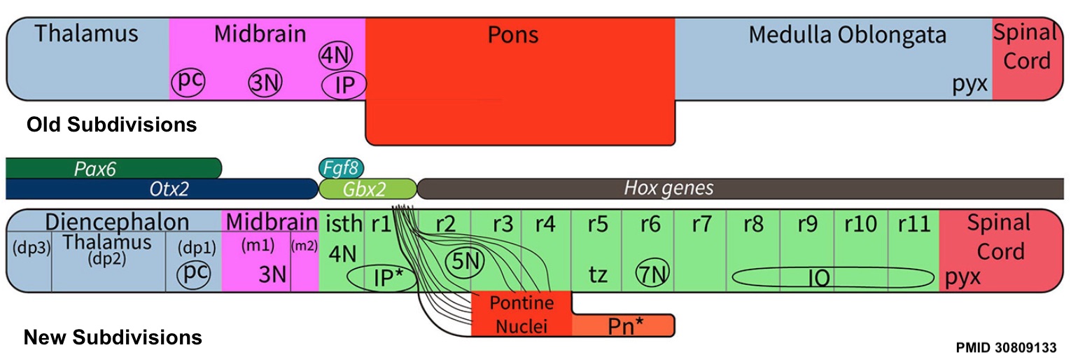
168 KB | Z8600021 | ==Comparison of brain stem subdivisions== the traditional view of subdivisions of the human brain stem with the new system of segmentation revealed by developmental gene expression. At the top, the subdivisions of the “old” human brain stem (the traditional version) are based on the assumption that the midbrain extends from the thalamus to the rostral margin of the pons; this concept wrongly holds that the pretectum (dp1) and the isthmus (isth) belong to the midbrain (Puelles et al., 2012a)... | 1 |
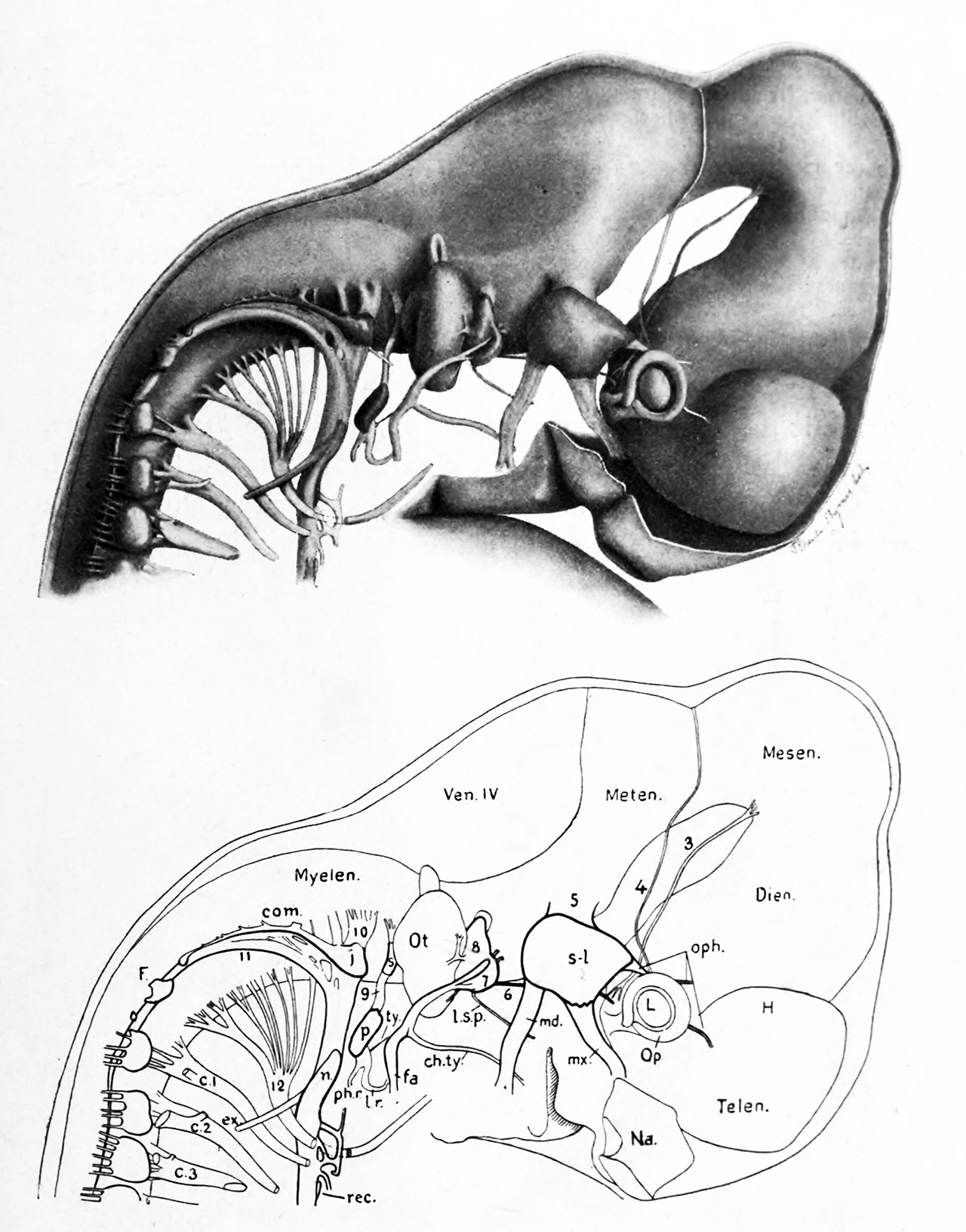
| 00:35, 24 February 2020 | Johnston1907 fig024.jpg (file) |  |
87 KB | Z8600021 | 3 | |
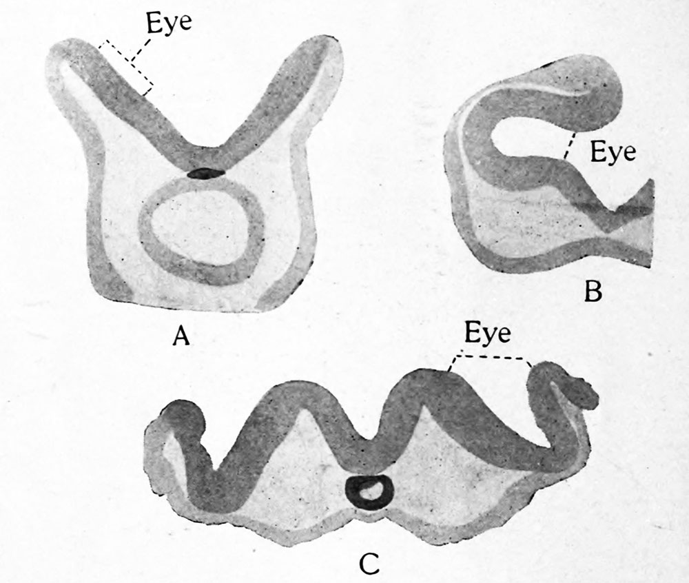
| 00:30, 24 February 2020 | Johnston1907 fig023.jpg (file) |  |
93 KB | Z8600021 | 3 | |
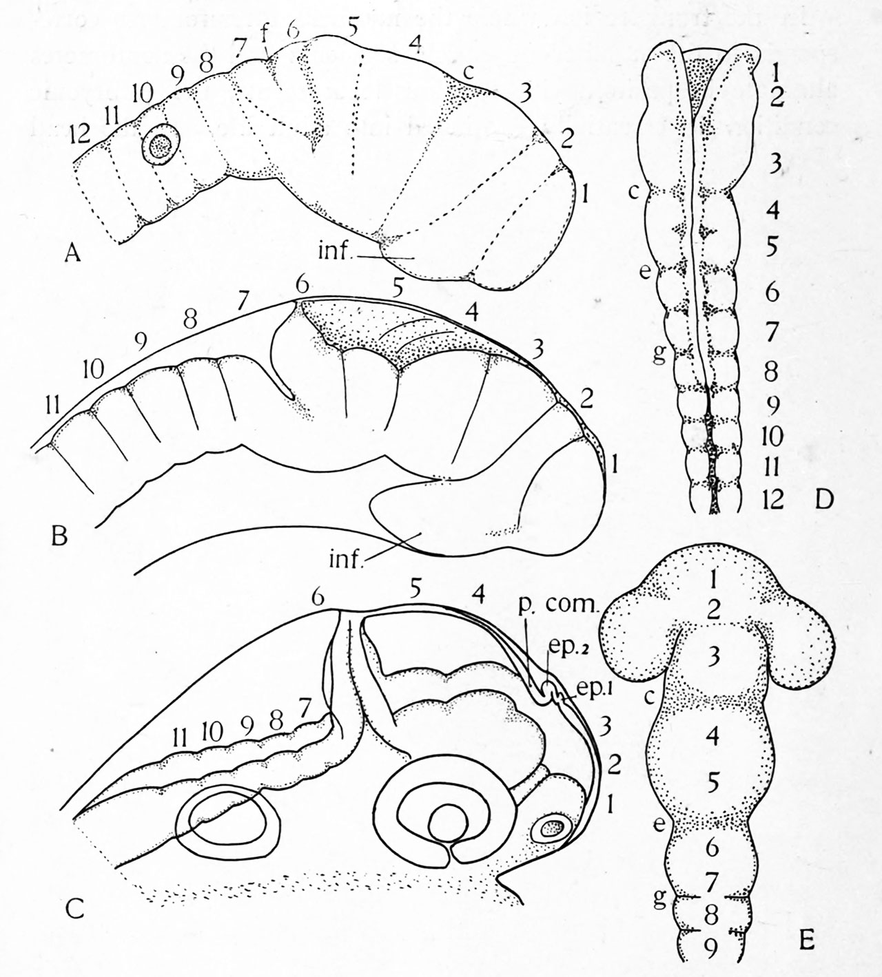
| 00:16, 24 February 2020 | Johnston1907 fig022.jpg (file) |  |
170 KB | Z8600021 | 3 | |
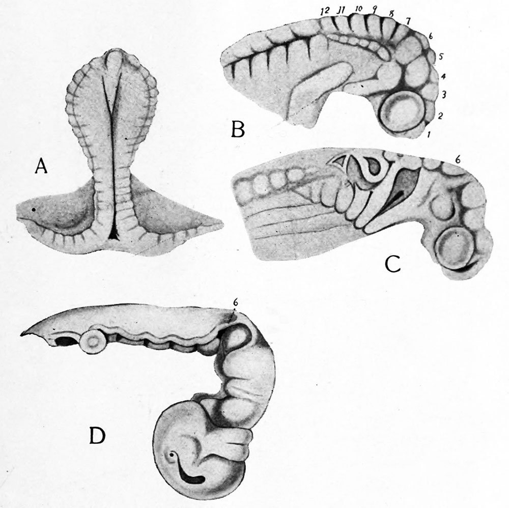
| 00:06, 24 February 2020 | Johnston1907 fig021.jpg (file) |  |
98 KB | Z8600021 | 2 | |
| 23:58, 23 February 2020 | Johnston1907 fig020.jpg (file) |  |
277 KB | Z8600021 | 2 | |
| 23:48, 23 February 2020 | Johnston1907 fig019.jpg (file) |  |
236 KB | Z8600021 | 2 | |
| 23:40, 23 February 2020 | Johnston1907 fig018.jpg (file) |  |
119 KB | Z8600021 | 2 | |
| 23:25, 23 February 2020 | Johnston1907 fig017.jpg (file) |  |
47 KB | Z8600021 | 2 | |
| 23:17, 23 February 2020 | Johnston1907 fig016.jpg (file) |  |
104 KB | Z8600021 | 2 | |
| 23:11, 23 February 2020 | Johnston1907 fig015.jpg (file) |  |
100 KB | Z8600021 | Reverted to version as of 23:06, 23 February 2020 (AEDT) | 4 |
| 22:58, 23 February 2020 | Johnston1907 fig014.jpg (file) |  |
69 KB | Z8600021 | 2 | |
| 22:51, 23 February 2020 | Johnston1907 fig013.jpg (file) |  |
135 KB | Z8600021 | 2 | |
| 22:39, 23 February 2020 | 1907 The Nervous System of Vertebrates titlepage.jpg (file) |  |
62 KB | Z8600021 | The Nervous System of Vertebrates (1907) {{Johnston1907 footer}} | 1 |
| 18:37, 23 February 2020 | 1907 Johnston - The nervous system of vertebrates.pdf (file) | |
25.69 MB | Z8600021 | {{Johnston1907 footer}} Category:PDF | 1 |
| 11:59, 20 February 2020 | Reference Handbook of the Medical Sciences titlepage.jpg (file) |  |
141 KB | Z8600021 | 2 | |
| 11:45, 20 February 2020 | 1901 Comparative development of the coelom.pdf (file) | |
5.49 MB | Z8600021 | =Comparative Development of the Coelom= ===Reference=== {{Ref-Mall1901coelom1}} {{Footer}} Category:Historic EmbryologyCategory:1900'sCategory:Coelomic Cavity Category:PDF | 1 |
| 13:01, 17 February 2020 | Pregnancy-care-guidelines-5Feb18-1.jpg (file) |  |
81 KB | Z8600021 | 2019 Edition cover | 2 |
| 10:07, 17 February 2020 | 2018 WHO diphtheria-tetanus-pertussis vaccine.jpg (file) |  |
259 KB | Z8600021 | ==2018 WHO - diphtheria-tetanus-pertussis vaccination== ===Reference=== [https://www.who.int/en/news-room/fact-sheets/detail/immunization-coverage WHO immunization coverage] | 1 |
| 19:29, 12 February 2020 | Carl G. Hartman.jpg (file) |  |
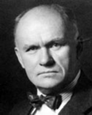
14 KB | Z8600021 | ==Carl G. Hartman (1879-1968)== Studied the opossum and rhesus monkey as models of the mammalian sexual cycles. A lab associate at the Laboratory of Embryology in 1925. :Links: Carl G. Hartman {{Footer}} Category:Historic EmbryologyCategory:1920'sCategory:PeopleCategory:USA | 1 |
| 18:46, 12 February 2020 | Carnegie Institution - Department of Embryology 1929.jpg (file) |  |
112 KB | Z8600021 | ==The Carnegie Institution of Washington - Department of Embryology (1929)== From left: Warren H Lewis, George L Streeter, Martin Nordmann, Emanuel Fauré-Fremiet, Zygmunt Grodzinski, RH Hunter, Chester H Heuser, Carl G Hartman and Otto Leif Tinklepaugh. ===Reference=== Photo - Alan Mason Chesney Medical Archives, Johns Hopkins Medical Institutions {{Footer}} | 1 |
| 12:09, 12 February 2020 | 1946 Development of the egg of the cow up to the stage of blastocyst formation.pdf (file) | |
4.62 MB | Z8600021 | Category:PDFCategory:Historic EmbryologyCategory:1940's | 1 |
| 10:43, 11 February 2020 | George John Romanes 1916-2014.jpg (file) |  |
145 KB | Z8600021 | ==George John Romanes (1916-2014)== Photograph - Romanes in 1984 {{Footer}} | 1 |
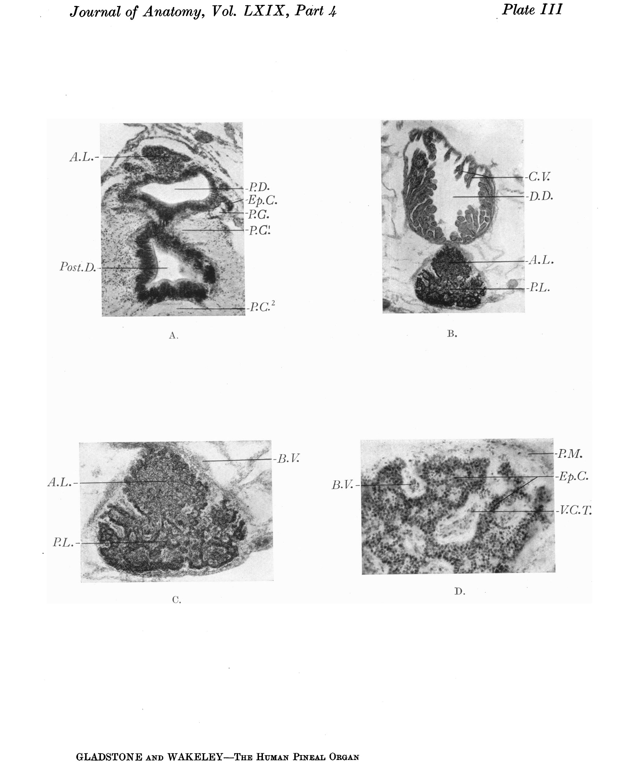
| 10:12, 10 February 2020 | Gladstone1935 plate3.jpg (file) |  |
574 KB | Z8600021 | {{Ref-Gladstone1935}} | 1 |
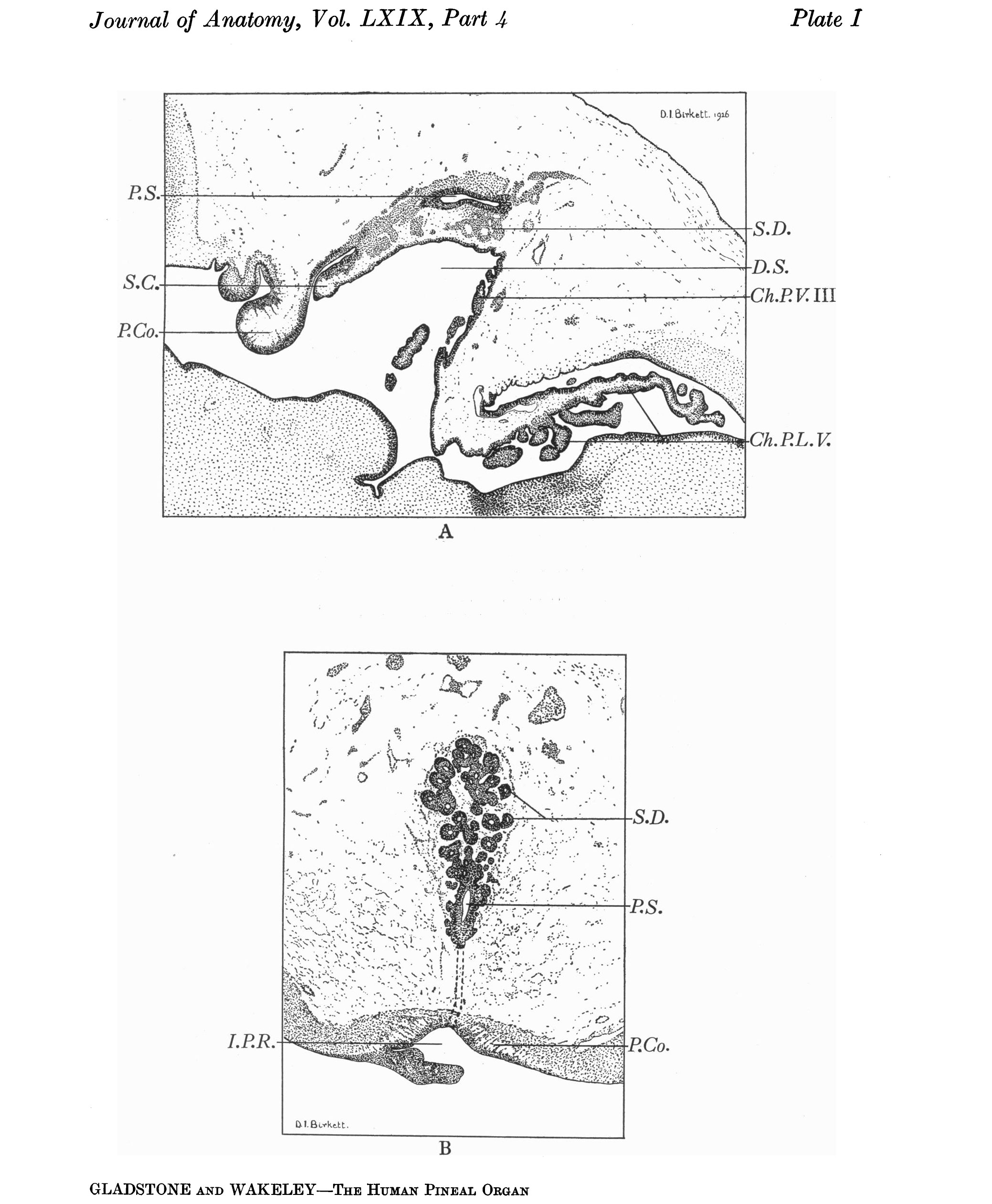
| 10:04, 10 February 2020 | Gladstone1935 plate1.jpg (file) |  |
541 KB | Z8600021 | {{Ref-Gladstone1935}} | 1 |
| 10:04, 10 February 2020 | Gladstone1935 plate2.jpg (file) |  |
972 KB | Z8600021 | {{Ref-Gladstone1935}} | 1 |
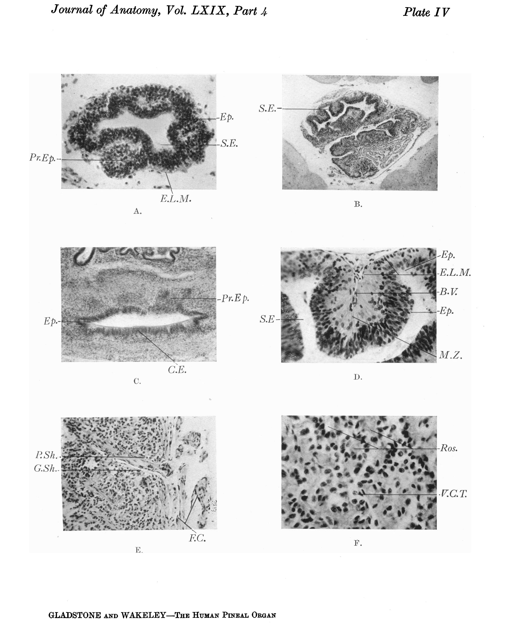
| 10:03, 10 February 2020 | Gladstone1935 plate4.jpg (file) |  |
804 KB | Z8600021 | {{Ref-Gladstone1935}} | 1 |
| 10:03, 10 February 2020 | Gladstone1935 plate5.jpg (file) |  |
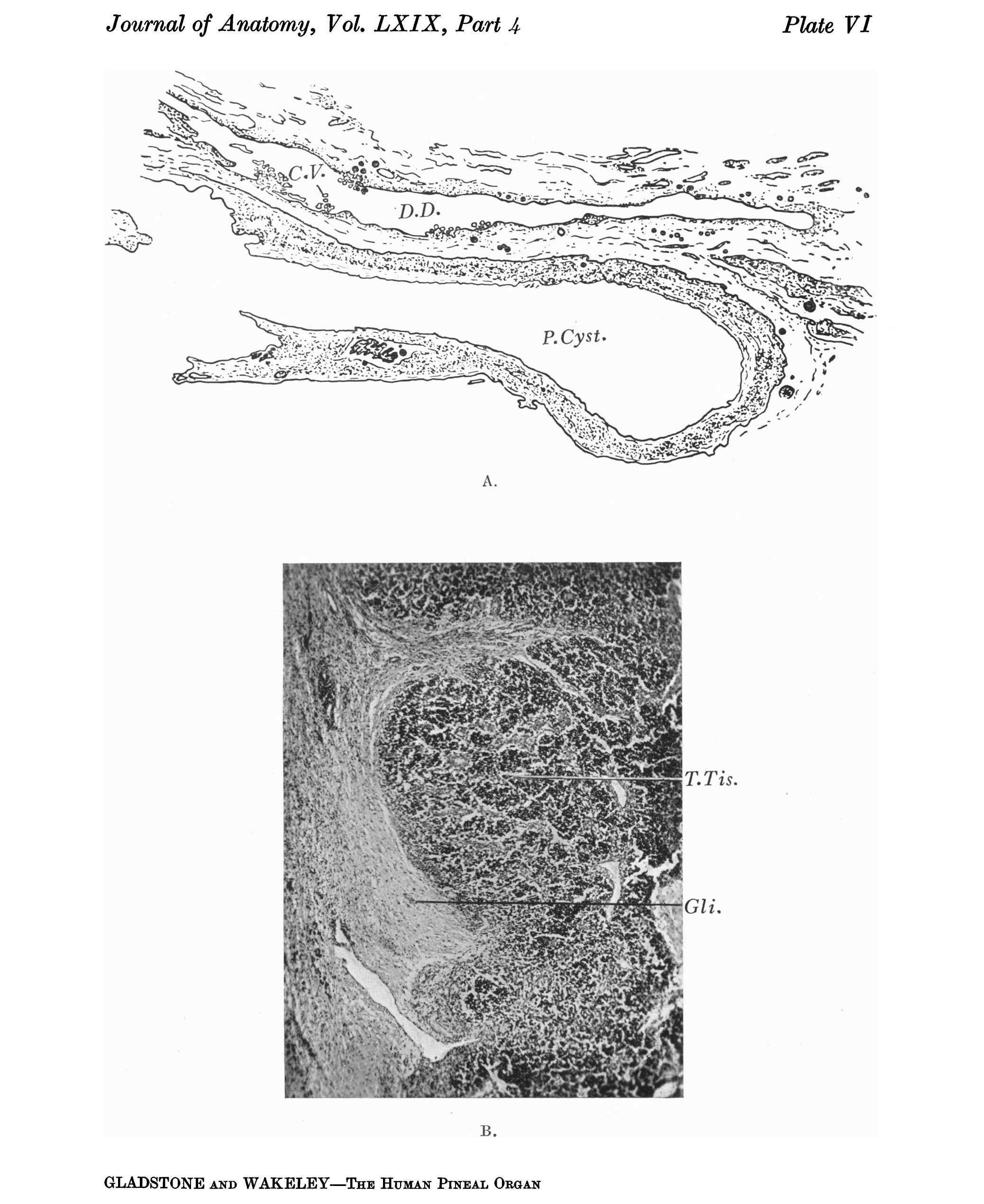
812 KB | Z8600021 | {{Ref-Gladstone1935}} | 1 |
| 10:03, 10 February 2020 | Gladstone1935 plate6.jpg (file) |  |
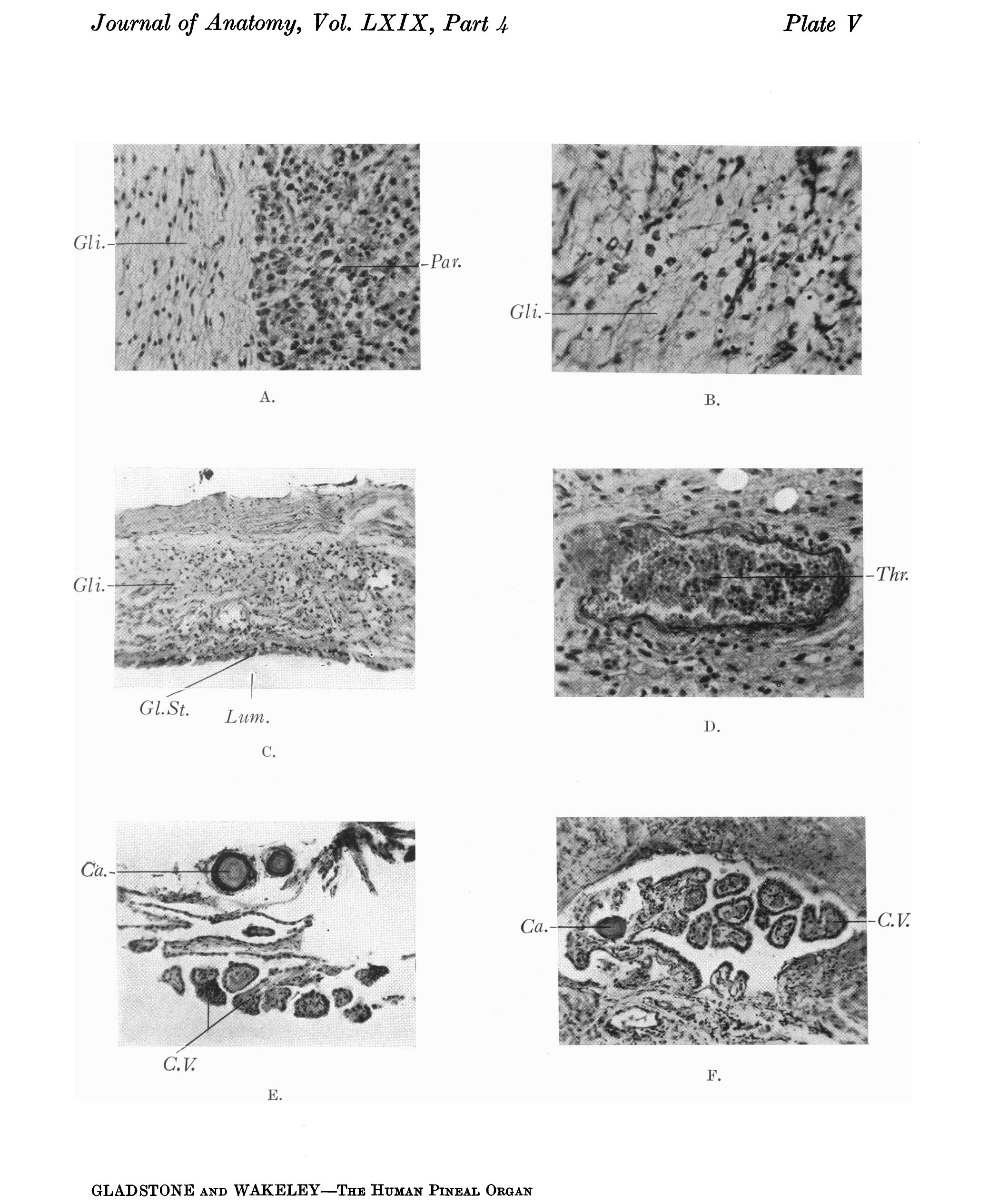
728 KB | Z8600021 | {{Ref-Gladstone1935}} | 1 |
| 11:50, 9 February 2020 | Flecker1932 fig10.jpg (file) |  |
53 KB | Z8600021 | 1 | |
| 11:50, 9 February 2020 | Flecker1932 fig09.jpg (file) |  |
23 KB | Z8600021 | 1 | |
| 11:49, 9 February 2020 | Flecker1932 fig08.jpg (file) |  |
18 KB | Z8600021 | 1 | |
| 11:49, 9 February 2020 | Flecker1932 fig07.jpg (file) |  |
58 KB | Z8600021 | 1 | |
| 11:49, 9 February 2020 | Flecker1932 fig06.jpg (file) |  |
106 KB | Z8600021 | 1 | |
| 00:00, 8 February 2020 | Flecker1932 fig05.jpg (file) |  |
47 KB | Z8600021 | 1 | |
| 23:59, 7 February 2020 | Flecker1932 fig04.jpg (file) |  |
41 KB | Z8600021 | 1 | |
| 23:59, 7 February 2020 | Flecker1932 fig03.jpg (file) |  |
44 KB | Z8600021 | 1 | |
| 23:59, 7 February 2020 | Flecker1932 fig02.jpg (file) |  |
41 KB | Z8600021 | 1 | |
| 23:59, 7 February 2020 | Flecker1932 fig01.jpg (file) |  |
42 KB | Z8600021 | 1 | |
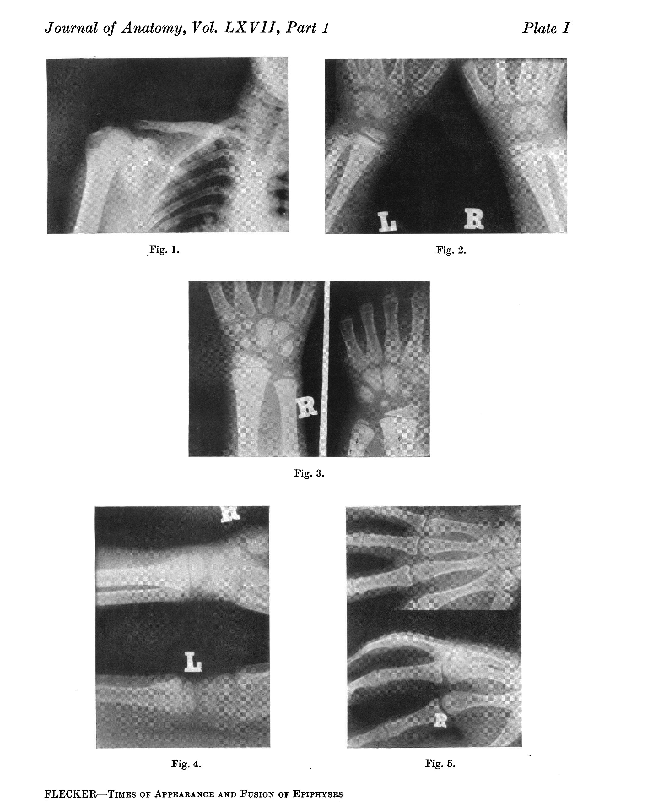
| 23:32, 7 February 2020 | Flecker1932 plate2.jpg (file) |  |
525 KB | Z8600021 | 1 | |
| 23:30, 7 February 2020 | Flecker1932 plate1.jpg (file) |  |
657 KB | Z8600021 | {{Ref-Flecker1932}} | 1 |
| 15:25, 2 February 2020 | Embryonic upper limb - brachial and superficial brachial artery.jpg (file) |  |
122 KB | Z8600021 | 2 | |
| 11:15, 2 February 2020 | Aortic arch branching pattern abnormalities.jpg (file) |  |
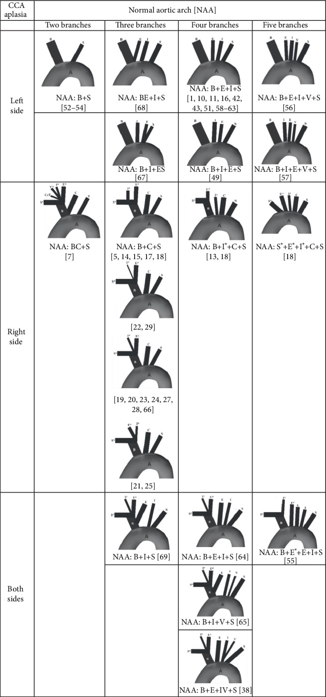
133 KB | Z8600021 | ==Aortic arch branching pattern abnormalities== Patterns of the branching of the normal aortic arch in cases of uni- and bilateral aplasia of the common carotid artery (CCA). Note: Kosinski, 1868 [66], cited by Boyd [14]. A, aorta; B, brachiocephalic trunk, S (S∗), left (right) subclavian artery; E (E∗), left (right) external carotid artery; I (I∗), left (right) internal carotid artery; V (V∗), left (right) vertebral artery; C, left common carotid artery; CcT, costocervical trunk. ===Refere... | 1 |