Search results
From Embryology
Page title matches
- [[Coelomic_Cavity_Development#Intra-embryonic_Coelom|coelom]]<noinclude>[[Category:Template]][[Category:Term Link]][[Category:Coelomic176 bytes (18 words) - 11:26, 25 January 2024
- [[Coelomic_Cavity_-_Abnormalities|coelom abnormalities]]<noinclude>[[Category:Template]][[Category:Term Link]][[Cate183 bytes (19 words) - 13:15, 24 January 2019
- [[Coelomic_Cavity_Development#Intra-embryonic_Coelom|intra-embryonic coelom]]<noinclude>[[Category:Template]][[Category:Term Link]][[Category:Coelomic269 bytes (26 words) - 10:53, 15 May 2019
- #REDIRECT [[Paper - Development of the human coelom (1897)]]60 bytes (8 words) - 10:04, 20 October 2020
- DEVELOPMENT OF THE HUMAN COELOM. ...h was emphasized the separation of the body cavity from the extraembryonic coelom. Since then I have had opportunity to extend my observation to the human em101 KB (17,581 words) - 14:15, 12 September 2017
- filled the various spaces of the coelom as they hloodlvessels ; Coc, coelom; Spl., splanchnopleure ; In., intestine; au.. aorta; Wd., Wolffian duct; Ye125 KB (20,812 words) - 09:59, 20 February 2020
- [[Media:1901 Comparative development of the coelom.pdf|Paper PDF]] =Development of the Human Coelom=102 KB (17,328 words) - 11:41, 20 February 2020
- ...s historic 1998 paper by Mall describes the early development of the human coelom, body cavity. Note "extraembryonic" appears in the paper without a hyphen, =Development of the Human Coelom=100 KB (17,375 words) - 10:03, 20 October 2020
- [[Media:1901 Comparative development of the coelom.pdf|Paper PDF]] =Comparative Development of the Coelom=26 KB (4,145 words) - 23:07, 14 April 2020
- ..._Hill.jpg|90px|left]] This historic 1913 paper by Jones describes the the coelom and the diaphragm. =The Functional History of the Coelom and the Diaphragm=86 KB (14,540 words) - 12:27, 28 January 2020
- conditions that prevail in animals which do not possess a coelom—the great The Functional History of the Coelom and the Diaphragm 28787 KB (14,781 words) - 11:12, 28 January 2020
Page text matches
- ...ence]][[Category:Franklin Mall]][[Category:Historic Embryology]][[Category:Coelom]][[Category:1800's]]</noinclude>299 bytes (33 words) - 09:57, 20 October 2020
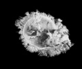
File:Mall1917 fig02.jpg ...o. 12S9 from Dr. J. R. Cottell, Louisville, Ky. X 2. The picture shows the coelom filled mostly with granular magma.(629 × 525 (29 KB)) - 18:31, 5 November 2013- | Extraembryonic Coelom | Intraembryonic Coelom588 bytes (62 words) - 08:13, 15 January 2019
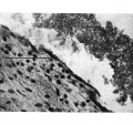
File:Wyburn1939-fig16.jpg ==Fig. 16. Photomicrograph of endothelial lining of umbilical cord coelom of embryo 42 mm== ...to cord “pillars” and continuous with rectus sheath. U.C. = umbilical cord coelom. L. = degenerating endothelial cells. Description in text.(848 × 800 (204 KB)) - 17:01, 14 September 2015- ...n coelom|1897 human coelom]] | [[Book - Manual of Human Embryology 13|1910 Coelom and Diaphragm]] | [[Paper - A stage in the development of the serous caviti449 bytes (57 words) - 09:14, 2 October 2020
File:Wyburn1939-fig15.jpg ==Fig. 15. Photomicrograph of section of umbilical cord coelom of embryo 42 mm== ...to cord “pillars” and continuous with rectus sheath. U.C. = umbilical cord coelom. L. = degenerating endothelial cells. Description in text.(848 × 800 (189 KB)) - 17:02, 14 September 2015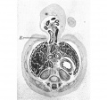
File:Wyburn1939-fig13.jpg Fig. 16. Photomicrograph of endothelial lining of umbilical cord coelom of embryo 42 mm. x circa 225. ...to cord “pillars” and continuous with rectus sheath. U.C. = umbilical cord coelom. L. = degenerating endothelial cells. Description in text.(848 × 800 (153 KB)) - 17:02, 14 September 2015- ...Handb. Med. Sci.}} 3: 171-189. [[Media:1901 Comparative development of the coelom.pdf|PDF]]<noinclude>[[Category:Template]][[Category:Reference]][[Category:C409 bytes (47 words) - 11:38, 20 February 2020
- ...ry of the coelom and the diaphragm (1913)|'''The functional history of the coelom and the diaphragm''']]. (1913) J Anat Physiol. 47(3):282-318. [https://www.414 bytes (50 words) - 11:06, 28 January 2020
- ...Handb. Med. Sci.}} 3: 166-171. [[Media:1901 Comparative development of the coelom.pdf|PDF]]<noinclude>[[Category:Template]][[Category:Reference]][[Category:C421 bytes (47 words) - 10:51, 20 February 2020
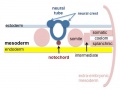
File:Mesoderm-cartoon4.jpg ...inar embryo showing the further development of the 3 layers and the space (coelom) that forms in the mesoderm (only the righthand side is shown, lefthand sid ...sion into somatic and {{splanchnic mesoderm}} separated by intra-embryonic coelom.(400 × 300 (20 KB)) - 10:09, 23 April 2020- F.PC.C. Floor of pericardial coelom. M.B. Medial boundary of pericardio-peritoneal coelom.1 KB (179 words) - 09:50, 9 October 2018
- #REDIRECT [[Paper - Development of the human coelom (1897)]]60 bytes (8 words) - 10:04, 20 October 2020
File:Wyburn1939-plate04.jpg Fig. 15. Photomicrograph of section of umbilical cord coelom of embryo 42 mm. Site indicated by arrow in Fig. 14. x circa 75. Fig. 16. Photomicrograph of endothelial lining of umbilical cord coelom of embryo 42 mm. x circa 225.(1,700 × 2,247 (747 KB)) - 09:38, 15 September 2015- ...le:Mesoderm-cartoon4.jpg|thumb|alt=Coelomic Cavity cartoon|Intra-embryonic coelom week 3-4 ({{GA}} 5-6)]] The intra-embryonic coelom ({{Coelomic cavity}}) forms within the lateral plate mesoderm early in embr2 KB (221 words) - 08:52, 25 January 2019
- ! width=300px|Extra-embryonic Coelom ! width=300px|Intra-embryonic Coelom631 bytes (78 words) - 13:07, 20 February 2020
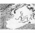
File:Wyburn1939-plate02.jpg ...I . =intercoelomic septum. D. = distal slit-like portion of umbilical cord coelom. S. = space in umbilical cord. U. = umbilical vein.(1,680 × 2,367 (786 KB)) - 17:13, 14 September 2015
File:Wyburn1939-text-fig08-10.jpg ...Horizontal lines = blood vessels. Black = mesoderm. U .C. = umbilical cord coelom. ...Horizontal lines = blood vessels. Black = mesoderm. U .C. = umbilical cord coelom.(1,588 × 1,400 (183 KB)) - 13:34, 15 September 2015- [[Coelomic_Cavity_Development#Intra-embryonic_Coelom|coelom]]<noinclude>[[Category:Template]][[Category:Term Link]][[Category:Coelomic176 bytes (18 words) - 11:26, 25 January 2024
- [[Coelomic_Cavity_-_Abnormalities|coelom abnormalities]]<noinclude>[[Category:Template]][[Category:Term Link]][[Cate183 bytes (19 words) - 13:15, 24 January 2019


