Search results
From Embryology
Page title matches
- ...ages that relate to the [[Carnegie Collection]] Embryo No. {{CE184}}. This embryo would be early fetal development [[Week 10]] based upon the CRL 50 mm. {{Carnegie Collection fetal table}}2 members (0 subcategories, 0 files) - 13:56, 18 November 2017
Page text matches
- ...e>[[Category:Template]][[Category:Carnegie Collection]][[Category:Carnegie Embryo]][[Category:Fetal]][[Category:1910's]]</noinclude>184 bytes (19 words) - 15:46, 7 November 2017
- ==Carnegie Fetus== Carnegie Embryo 13183 KB (314 words) - 12:24, 18 November 2017
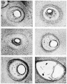
- ...gie Collection Embryo No. {{CE7950}}. This embryo was classified as late [[Carnegie stage 5|Stage 5]] occurring during [[Week 2]]. ...onic disc, 0.16 x 0.07 mm. Slightly more developed than No. 7699, although embryo is a little less differentiated. Photomicrographs in Hertig, Rock, and Adam2 members (0 subcategories, 1 file) - 17:59, 1 October 2018
- ...ages that relate to the [[Carnegie Collection]] Embryo No. {{CE184}}. This embryo would be early fetal development [[Week 10]] based upon the CRL 50 mm. {{Carnegie Collection fetal table}}2 members (0 subcategories, 0 files) - 13:56, 18 November 2017
- ==Embryo List== ! colspan=10| [[Carnegie Collection]] _ [[Carnegie stage 5|Stage 5]] 5 KB (696 words) - 10:21, 4 October 2018
- ! colspan=10| [[Carnegie Collection]] - [[Carnegie stage 5|Stage 5]] ...age, having a developmental age of about eight and nine days respectively. Carnegie Instn. Wash. Publ. 583, [[Book - Contributions to Embryology|Contrib. Embry5 KB (585 words) - 16:15, 24 October 2017
- * [[:Category:Florian Embryo Bi II|Florian Embryo Bi II]] 4–5 somites [[Carnegie stage 10]] * [[:Category:Florian Embryo Bi III|Florian Embryo Bi III]] 4–5 somites [[Carnegie stage 10]]3 KB (439 words) - 11:48, 8 February 2020
File:Streeter014-19.jpg ...the lateral semicircular canal in a human fetu.s 43 mm. crown-rump length (Carnegie Collection, No. 886, slide 42, section 3). The section is 100m thick and is ...the lateral semicircular canal in a human fetus 46 mm. crown-rump length (Carnegie Collection, No. 9.5, slide 72, section 1). The section i.s 100m thick and i(640 × 800 (199 KB)) - 02:46, 15 February 2011- [[Image:Stage9sm.jpg|thumb|Carnegie stage 9 showing somite formation]] [[Image:Stage 9 SEM1.jpg|thumb|Carnegie stage 9 scanning electron microscope image showing somite formation]]5 KB (773 words) - 12:04, 9 August 2010
- {{Carnegie No.20 Header}} Frontal section through the region of the ear in a human embryo 4 mm. long (Carnegie Collection, No. 588, slide 6, row 6, section 6). The section is 15 um thick18 KB (2,849 words) - 22:04, 22 April 2012
- [[Carnegie Collection]]: {{CE45}}, {{CE75}}, {{CE86}}, {{CE95}} ...en Kautenhirns''"; a description of these is accordingly unnecessary. Each embryo has been studied in serial sections and from tliese sections a few, at diff17 KB (2,866 words) - 04:19, 19 February 2020
- {{Carnegie No.20 Header}} ...0 mm. embryo by compared with a similar cast of the same canal in a 30 mm. embryo, it will be seen that the general form of the canal in the older specimen i25 KB (3,995 words) - 08:59, 28 August 2011
- ...blood development, including the fact that the red corpuscles in the early embryo are nucleated and that the later cells lack a nucleus. Specific informatio ...red blood cells begins to be evident during the second month in the human embryo and (2) that few nucleated red cells are found by the middle of the third m13 KB (2,081 words) - 21:13, 31 May 2018
- [[Image:Stage9sm.jpg|thumb|Carnegie stage 9 showing somite formation]] [[Image:Stage 9 SEM1.jpg|thumb|Carnegie stage 9 scanning electron microscope image showing somite formation]]16 KB (2,146 words) - 01:53, 28 August 2010
- ...Mall describes the human embryos in the collection that would become the [[Carnegie Collection]]. There is also a [[:File:1904 - Catalogue of the collection of [[Carnegie Collection]] | [[Carnegie Embryos]]21 KB (2,470 words) - 23:39, 9 August 2018
- ...storic 1944 paper by Davies describes early human development in week 2 ([[Carnegie stage 5]]). {{Carnegie stage 5 links}}20 KB (3,201 words) - 17:47, 1 October 2018
- ...c 1957 paper by O'Rahilly is a description of the development of the human embryo limb cartilage. ...4; Hagen, 1900; Lewis, 1902; Griifenberg, I905; Hesser, 1926). In a 27—mm. embryo, Schulin (1879) found that all the skeletal elements of the hand were ehond43 KB (6,197 words) - 07:54, 29 April 2017
- ...istoric histological study of the development of the meninges of the human embryo spinal cord. Our current understanding of interstitial cell development and ...by the Department of Embryology, sincere appreciation is expressed to the Carnegie Institution of Washington, and to Dr. G. W. Corner, Director. The author is49 KB (7,379 words) - 12:44, 25 July 2020
- ...e free to use our judgment in methods of fixation and preservation. If the embryo is perfectly fresh or possibly living, we use, of course, the most refined ...straight and other measurements and weights also are taken. The age of the embryo is estimated on the basis of weight, crown-rump, and foot length, and the e56 KB (7,365 words) - 04:08, 19 February 2020
- ...e will cover the early development of the ectoderm layer of the trilaminar embryo. Note that we will be returning later to discuss neural (central nervous sy ...imation shows early neural development from week 3 onward. The whole early embryo development (dorsolateral view) is shown, yolk sac to left.41 KB (5,663 words) - 12:05, 4 October 2011
