User:Z3290841
-Z3290841 09:37, 30 July 2011 (EST)
Lab 1 Assessment
Identify the origin of In Vitro Fertilization (IVF) and the 2010 nobel prize winner associated with this technique
Origin of IVF
Studies and experiments about IVF have been going for more than a hundred years now. The first experiments and studies could date as far back as 1878, which involved the fertilisation of mammalian eggs in vitro, but were all unsuccessful. The source of this problem is the lack of knowledge about the need for egg cell maturation, sperm cell capacitation, and the correct media and condition for fertilisation. It was not until Austin and Chang coined and postulated sperm capacitaion that the technology produce more successful results.
Robert Edwards
He was the Nobel Prize laureate in 2010 in Physiology or Medicine for his contribution to the development of in vitro fertilisation. His work involved the development of a human culture media that allowed fertilisation and early embryo culture.
Identify a recent paper on fertilisation and describe its key findings
Terada Y, Hasegawa H, Ugajin T, Murakami T, Yaegashi N, Okamura K.(2009).Microtubule organization during human parthenogenesis.Fertility and Sterility, 91(4), 1271-2. Retrieved from http://www.ncbi.nlm.nih.gov/pubmed/18706544
The key finding of the experiment that they did was the presence of multiple microtubule organising centre (MTOC) in human oocyte cytoplasm during parthenogenesis, which was previously only known to have been produced by the sperm centrosome in human fertilisation.
Identify 2 congenital anomalies
- Arterial Septal Defect - type of congenital heart defect, whereby the blood flow between the left and right atria is through the interatrial septum
- Spina Bifida - type of developmental congenital disorder, whereby there is an incomplete closing of the embryonic neural tube.
--Mark Hill 09:57, 3 August 2011 (EST) These answers are fine.
--z3290841 11:52, 4 August 2011 (EST)
Lab 2 Assessment
Identify the ZP protein that spermatozoa binds and how is this changed (altered) after fertilisation
Zona Pellucida Protein 3 (ZP3) is the glycoprotein where spermatozoa binds to, triggering an acrosome reaction that releases lytic enzymes, like acrosin and glycosidases. This aids in the movement of the sperm through Zona Pellucida. The ZP3 protein losses its ability to induce acrosome reaction and receive sperms once fertilisation occurs.
I did research on Cystic fibrosis and these are the 2 articles that I found that explains a lot about the disease, and the genetic research on it.
Review Article:
Cystic Fibrosis: Seminar
Description: This is basically outlining the pathophysiology of the disease, disease manifestation,current treatments diagnostic tool.
Ratjen F & Doring G.(2003).Cystic Fibrosis: Seminar.The Lancet, 361, 681-9. Retrieved from http://www.sciencedirect.com/science/article/pii/S0140673603125676
Research Article:
Gene expression profile study in CFTR mutated bronchial cell lines
Description: This article provides information about the severity of gene expression in relation of the extent or type of mutation.
Gambardella S, Biancolella M, D'Apice M, Amati F, Sangiuolo F, Farcomenti A, Chillemi G, Bueno S, Desideri A & Novelli G.(2006).Gene expression profile study in CFTR mutated bronchial cell lines.Clinical and Experimental Medicine , 6, 157-65. Retrieved from http://proquest.umi.com/pqdlink?Ver=1&Exp=08-05-2016&FMT=7&DID=1187181501&RQT=309&cfc=1
--z3290841 20:05, 10 August 2011 (EST)
--z3290841 11:10, 11 August 2011 (EST)
Lab 3 Assessment
What is the maternal dietary requirement for late neural development
Iodine
Dietary Requirement = 0.22 mg daily
Food Source = Seafood and iodized salt
Role = Important in the production of maternal thyroid hormone, which is not only needed by the mother but also by the developing fetus. This is because of its role in neuronal migration and myelination. Lack of iodine, thus thyroid hormone, may lead to cretinism, which is a syndrome characterised by permanent brain damage and mental retardation.
For more information see: http://www.pronutrition.org/files/MaternalNutritionDietaryGuide.pdf
Omega-3 fatty acid DHA
Dietary Requirement > 300 mg daily
Food Source = pink salmon, white tuna
Role = an essential fatty acid that promotes neural stem cell differentiation into neurons. This is by prompting cell cycle exit and suppressing cell death
Folic Acid
Dietary Requirement = 0.4 mg daily
Food source = dark green leafy vegetables, breakfast cereals, beans (legumes), whole grains
Role = Consumption reduces the risk of infant neural tube defects because it is an essential component for producing follate which is needed in DNA synthesis.
Zinc
Dietary Requirement > 11 mg daily
Food Source = Organ meats, red meat, poultry, whole fish
Role = It is said to have a role in the absorption of folic acid, thus lack of it may also lead to neural tube defects.
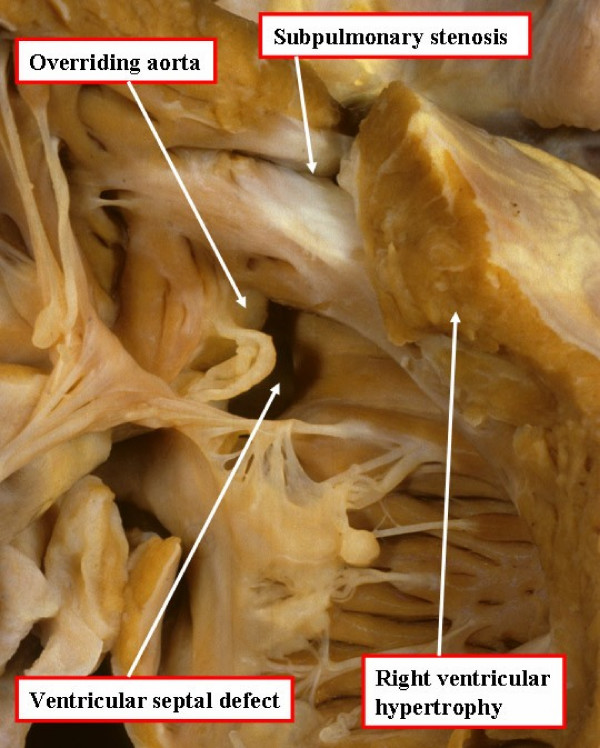
Upload a picture relating to you group project
Tetralogy of Fallot
--z3290841 11:14, 15 August 2011 (EST)
--z3290841 12:59, 18 August 2011 (EST)
Lab 4 Assessment
The allantois, identified in the placental cord, is continuous with what anatomical structure
The allantois arises from the posterior part of the yolk sac. After the development of the hind-gut, around week 4-5, it is continuous with the cloaca, which is the terminal part of the hind-gut. It also moves closer to the umbilical vessels and becomes part of the umbilical cord.
Identify the 3 vascular shunts, and their location, in the embryonic circulation
- Ductus Venosus - connects the portal and umbilical veins to the inferior vena cava
- Ductus Arteriosus - connects pulmonary trunk to aorta
- Foramen Ovale - connects right and left atrium
Identify the Group project sub-section that you will be researching
- Introduction
- History
- Epidemiology
- Signs and Symptoms
- Genetics/Aetiology
- Pathophysiology and Abnormalities
- Diagnostic Tests
- Treatment/Management
- Prognosis
- Future Directions
- References
--z3290841 08:40, 24 August 2011 (EST)
--z3290841 12:49, 25 August 2011 (EST)
Lab 5 Assessment
Which side (L/R) is most common for diaphragmatic hernia and why
One of the developmental abnormalities of the diaphragm is Congenital Diaphragmatic Hernia (CDH), and a left sided Bochdalek hernia is the most common type of the abnormality. This means that the herniation occurs at the postero-lateral area of the diaphragm. The early closure of the right pluroperitoneal opening might be the cause as to why a left-sided CDH is more common.
--z3290841 08:20, 1 September 2011 (EST)
--z3290841 11:22, 1 September 2011 (EST)
Lab 6 Assessment
What week of development do the palatal shelves fuse
The palatal shelves fuse during the 9th Week of embryonic development. This involves the growth, elevation and fusion of primary palate, and then fusion with the secondary palate[1].
What early animal model helped elucidate the neural crest origin and migration of neural crest cells
The quail-chick chimera was the early animal model used by Nicole Le Douarin to elucidate this. This is by grafting a neural crest tube and cells in the host chick's embryo. This is because Le Dourin have identified that a stain called the Fuelgen stain is ale to distinguish the cell form quail and chick[2].
What abnormality results from neural crest not migrating into the cardiac outflow tract
Some of these abnormalities include:
- Persistent Truncus Arteriosus (PTA)
- Double outlet right ventricle (DORV)
- Dextraposed aorta (DA)
- Tetralogy of Fallot (TOF)
- Ventricle septum defect (VSD)
Eperiments on mouse showed that partial ablation of cardiac neural crest results to DORV, DA, TOF and VSD, while PTA is the result of complete ablation of cardiac neural crest[3].
--z3290841 08:21, 15 September 2011 (EST)
--z3290841 11:08, 15 September 2011 (EST)
Lab 7 Assessment
Are satellite cells (a) necessary for muscle hypertrophy and (b) generally involved in hypertrophy
According to studies on satellite cells, they are not necessary for muscle hypertrophy, but they still undergo hypertrophy. This is why in the experimental studies of skeletal muscle the results showed that the muscle being studied underwent hypertrophy even though satellite cells have been deactivated. At the same time in another experiment where satellite cells haven't been deactivated, the muscle cells have increased in number, which is double the amount compared to when satellite cells are deactivated. This illustrates the ability of satellite cells to hypertrophy.
Why does chronic low frequency stimulation cause a fast to slow fibre type shift
Chronic low frequency stimulation mimics the slow electrical signalling of motor neurons. Slow fibre muscles are more sensitive and responsive to these types of signals. Studies conducted investigating types of muscle fibres and conversion of the fibres to different types illustrated that these electrical stimulation/signals have the ability to transform/convert fast muscle fibres to slow muscle fibres. This is because chronic stimulation of fast fibres by low frequency signals activates an adaptive response that converts the muscle fibre, but the conversion follows the "next neighbour" rule, whereby the muscle fibres get converted in stages from a fast muscle fibre type to a slow muscle fibre type. It follows the following sequence of fibre types, from fastest to slowest type:
- IIb
- IIx
- IIa
- I
Also note that it could go from slow to fast fibres as well.
Trisomy 21 page Evaluation
Good brief and concise introduction. The use of the links at the end of the introduction is very helpful as the reader doesn't have to go back and fort if a section for external links was just made, but I believe there are too many links to put in one section, especially the introduction, it makes the section too cluttered and busy. I believe some of the links could be reevaluated and see if they should be incorporated in the page/section.
The recent findings section was an amazing idea, as it allows the reader to see that there are current progress on the disease, and it also gives credibility to the page as being up-to-date with the information.
I believe the karyotyping, aneuploidy, and Meiosis 1 & 2 section can be combined together as 1 subheading talking about the genetics/aetiology of the disease, which is not really explored in the page. It would have been much more informative if the page discussed about the current theories as to how the aneuploidy occurred, and use the Meiosis 1 & 2 section as back up evidence for it. Basically some of the sections are informative but not really necessary, like the Aneuploidy section, and it would have been better if the genetics side of things have been further explored.
The associated congenital abnormalities, heart defect and limb defect could have been synthesised together and presented in a much more conducive way, where the information is organised in a more orderly fashion, like the use of subheadings. The information contained in each of the 3 sections mentioned above have enough detail to get an idea about the defects in the disorder.
The section American College of Obstetricians and Gynecologists Recommendations and Trisomy growth charts could be interchanged, as it may allow the page to flow better than how it is.
The section of Prevalence and Diagnosis are done very well and are very informative.
The images and diagrams are used effectively throughout the whole page, there are no diagrams that should not be found in a section that it should not be in.
Overall it is a good page, not outstanding. It could still use some more information in it and the structuring could still be modified.
--z3290841 08:33, 22 September 2011 (EST) --z3290841 11:58, 22 September 2011 (EST)

