File:Merkel cell EM 01.jpg

Original file (984 × 738 pixels, file size: 209 KB, MIME type: image/jpeg)
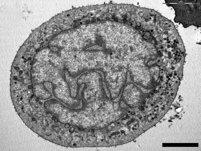
Electron microscopy analysis confirmed the identity of Merkel cells
Ultrastructural analyses were carried out on cells from the enriched MC fraction. (a, b) Up to half of the cells presented features characteristic of MCs: a polylobulated nucleus with numerous typical dense-core granules in a clear cytoplasm.
Scale bar 5 µm
Original file name: Figure 3. (panel a cropped from full figure, label removed and resized)
Reference
<pubmed>19668696</pubmed>| PLoS One.
Citation: Boulais N, Pereira U, Lebonvallet N, Gobin E, Dorange G, et al. (2009) Merkel Cells as Putative Regulatory Cells in Skin Disorders: An In Vitro Study. PLoS ONE 4(8): e6528. doi:10.1371/journal.pone.0006528
Copyright: © 2009 Boulais et al. This is an open-access article distributed under the terms of the Creative Commons Attribution License, which permits unrestricted use, distribution, and reproduction in any medium, provided the original author and source are credited.
File history
Yi efo/eka'e gwa ebo wo le nyangagi wuncin ye kamina wunga tinya nan
| Gwalagizhi | Nyangagi | Dimensions | User | Comment | |
|---|---|---|---|---|---|
| current | 18:10, 6 Haziran 2011 | | 984 × 738 (209 KB) | S8600021 (talk | contribs) | ==Electron microscopy analysis confirmed the identity of Merkel cells== Ultrastructural analyses were carried out on cells from the enriched MC fraction. (a, b) Up to half of the cells presented features characteristic of MCs: a polylobulated nucleus wit |
You cannot overwrite this file.
File usage
The following page uses this file: