File:HamiltonBoyd1960 fig02.jpg
From Embryology
Size of this preview: 574 × 600 pixels.
Original file (800 × 836 pixels, file size: 121 KB, MIME type: image/jpeg)
Summary
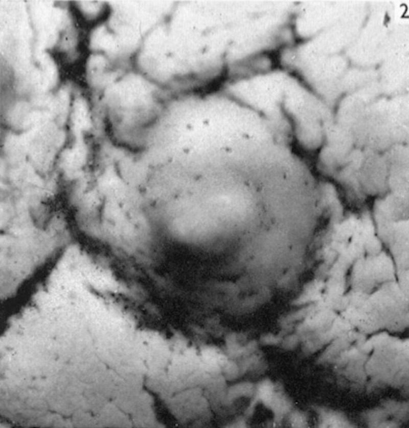
Fig. 2. Barnes Embryo
Photograph ( x 12) of the surface view of the implantation site of the Barnes embryo.
The implantation site itself appears as a slightly elevated area of the endometrium on which openings of the uterine glands can be seen. The surface of the endometrium shows characteristic shallow and irregular furrows.
File history
Yi efo/eka'e gwa ebo wo le nyangagi wuncin ye kamina wunga tinya nan
| Gwalagizhi | Nyangagi | Dimensions | User | Comment | |
|---|---|---|---|---|---|
| current | 13:11, 6 August 2020 | | 800 × 836 (121 KB) | Z8600021 (talk | contribs) | ==Fig. 2. Barnes Embryo== Photograph ( x 12) of the surface view of the implantation site of the Barnes embryo. The implantation site itself appears as a slightly elevated area of the endometrium on which openings of the uterine glands can be seen. The surface of the endometrium shows characteristic shallow and irregular furrows. |
You cannot overwrite this file.
File usage
The following 2 pages use this file: