File:HamiltonBoyd1960 plate04.jpg
Original file (1,280 × 1,710 pixels, file size: 515 KB, MIME type: image/jpeg)
Plate 4
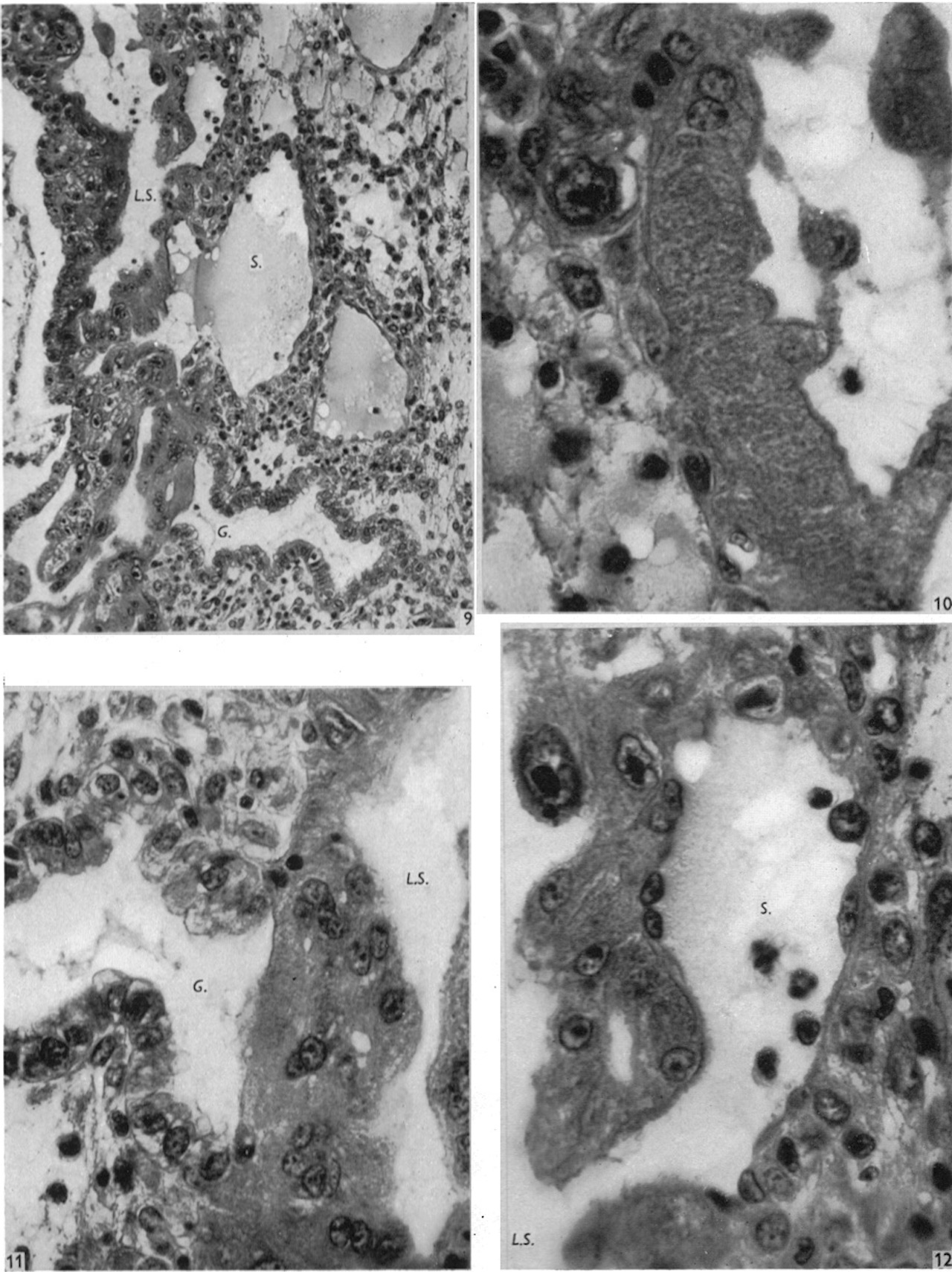
Fig. 9. Photograph ( x 200) of a section through the deep central portion of the implantation site of the Barnes embryo; maternal tissue is on the right of the illustration. The intereommunicating lacunar spaces (L.S.) are shown surrounded by syncytial trophoblast. Uterine venous sinusoids (S.) communicate with these. A plug of syncytium can be seen in direct contact with the lumen of the uterine gland (G.).
Fig. 10. Photomicrograph (x 840) of a section through the primitive syncytium and adjacent oedematous endometrium in the Barnes embryo; maternal tissue is on the left of the illustration. Note the texture of the syncytial cytoplasm, a long stretch of which is devoid of nuclei. In the upper part of the figure there is an irregular projection of syncytium, containing a very large nucleus, into the endometrium; elsewhere the foetal-maternal junction is smooth.
Fig. 11. Photomicrograph ( x 636) of the foetal-maternal junction of the Barnes embryo. A mass of primitive syncytium is in direct contact with the lumen of a uterine gland (G.) in which secretion can be seen. The syncytium separates this lumen from a lacunar space (L.S.).
Fig. 12. Photomicrograph ( x 840) to show an opening of a venous sinusoid (S.) into a lacunar space (L.S.). Note the relations of the endothelium of the sinusoid to the syncytium.
File history
Yi efo/eka'e gwa ebo wo le nyangagi wuncin ye kamina wunga tinya nan
| Gwalagizhi | Nyangagi | Dimensions | User | Comment | |
|---|---|---|---|---|---|
| current | 13:34, 6 August 2020 | | 1,280 × 1,710 (515 KB) | Z8600021 (talk | contribs) | crop, adjust size |
| 13:32, 6 August 2020 |  | 1,030 × 1,429 (365 KB) | Z8600021 (talk | contribs) |
You cannot overwrite this file.
File usage
The following 12 pages use this file:
- Paper - Development of the human placenta in the first three months of gestation (1960)
- File:HamiltonBoyd1960 fig02.jpg
- File:HamiltonBoyd1960 fig03.jpg
- File:HamiltonBoyd1960 fig04.jpg
- File:HamiltonBoyd1960 fig05.jpg
- File:HamiltonBoyd1960 fig06.jpg
- File:HamiltonBoyd1960 fig07.jpg
- File:HamiltonBoyd1960 fig08.jpg
- File:HamiltonBoyd1960 plate02.jpg
- File:HamiltonBoyd1960 plate03.jpg
- File:HamiltonBoyd1960 plate13.jpg
- Template:HamiltonBoyd1960 plates


