File:Lockwood1888b fig49.jpg
From Embryology
No higher resolution available.
Lockwood1888b_fig49.jpg (800 × 496 pixels, file size: 69 KB, MIME type: image/jpeg)
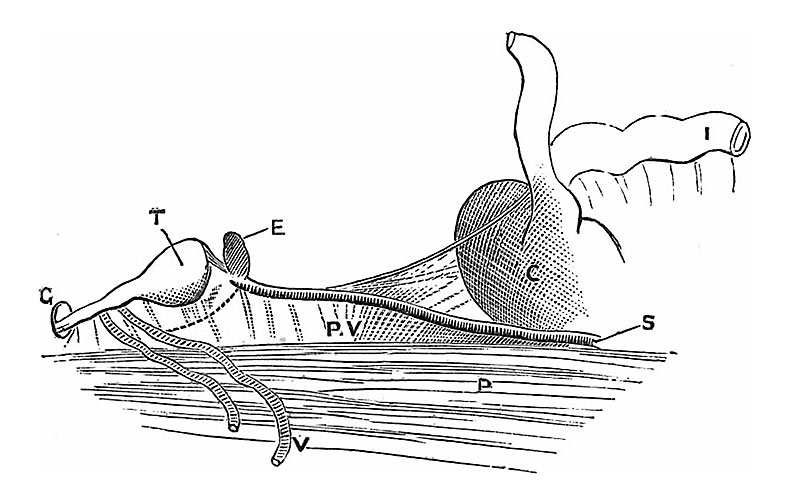
Fig. 49 — Drawing made from a seven or eight months’ foetus to show the fold (plica vascularis) which connects the testis with the cecum. T, testicles ; E, epididymis; P, psoas; V, vas deferens; G, plica gubernatrix, disappearing into processus vaginalis; P.V, plica vascularis ; C, cecum ; S,"spermatic artery ; I, ilium.
File history
Yi efo/eka'e gwa ebo wo le nyangagi wuncin ye kamina wunga tinya nan
| Gwalagizhi | Nyangagi | Dimensions | User | Comment | |
|---|---|---|---|---|---|
| current | 18:10, 14 April 2020 | | 800 × 496 (69 KB) | Z8600021 (talk | contribs) | adjust size W 800px |
| 18:10, 14 April 2020 |  | 927 × 575 (82 KB) | Z8600021 (talk | contribs) | crop | |
| 18:09, 14 April 2020 |  | 1,213 × 799 (136 KB) | Z8600021 (talk | contribs) |
You cannot overwrite this file.
File usage
The following page uses this file:


