File:Flint1906 plate01.jpg

Original file (1,280 × 843 pixels, file size: 149 KB, MIME type: image/jpeg)
Summary
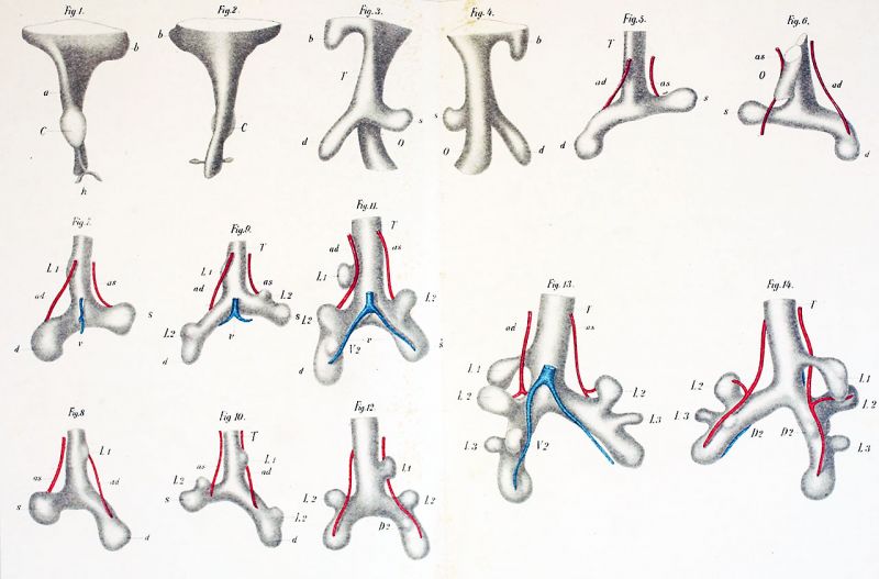
Plate I
figs. 1-14. are magnified 50 diameters. Pulmonary arteries red, pulmonary veins blue, bronchi white.
fiG. 1. Reconstruction of a portion of the head gut of a pig’s embryo 3 mm. long. Ventral view.
fiG. 2. Dorsal View of the same reconstruction.
fiG. 3. Reconstruction of a. portion of the head gut of a. pig's embryo 5 mm. long. Ventral view.
fiG. 4. Dorsal view of the same reconstruction.
fiG. 5 Reconstruction of the bronchial tree of a pig 6 mm. long.
fiG. 6. Dorsal view of the same reconstruction.
fiG. 7 Reconstruction of the bronchial tree of a. pig 7.5 mm. long.
fiG. 8 Dorsal View of the same reconstruction.
fiG. 9. Reconstruction of the bronchial tree of a. pig 8.5 mm. long.
fiG. 10. Dorsal View of the same reconstruction.
fiG. 11. Reconstruction of the bronchial tree of a pig 10 mm. long.
fiG. 12. Dorsal view of the same reconstruction.
fiG. 13. Reconstruction of the bronchial tree of a pig 12 mm. long.
fiG. 14. Dorsal view of the same reconstruction.
File history
Yi efo/eka'e gwa ebo wo le nyangagi wuncin ye kamina wunga tinya nan
| Gwalagizhi | Nyangagi | Dimensions | User | Comment | |
|---|---|---|---|---|---|
| current | 15:17, 8 April 2020 | | 1,280 × 843 (149 KB) | Z8600021 (talk | contribs) | adjust size, sharpness |
| 15:16, 8 April 2020 |  | 3,197 × 2,106 (531 KB) | Z8600021 (talk | contribs) | adjust colours | |
| 15:14, 8 April 2020 |  | 3,549 × 2,699 (525 KB) | Z8600021 (talk | contribs) | ==Plate I== figs. 1-14. fiGS. 1-20 are magnified 50 diameters. Pulmonary arteries red, pulmonary veins blue, bronchi white. fiG. 1. Reconstruction of a portion of the head gut of a pig’s embryo 3 mm. long. Ventral view. fiG. 2. Dorsal View of the same reconstruction. fiG. 3. Reconstruction of a. portion of the head gut of a. pig's embryo 5 mm. long. Ventral view. fiG. 4. Dorsal view of the same reconstruction. fiG. 5 Reconstruction of the bronchial tree of a pig 6 mm. long. fiG.... |
You cannot overwrite this file.
File usage
The following page uses this file: