File:Johnston1907 fig016.jpg
From Embryology
Size of this preview: 547 × 599 pixels. Other resolution: 730 × 800 pixels.
Original file (730 × 800 pixels, file size: 104 KB, MIME type: image/jpeg)
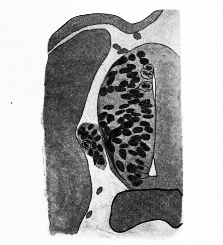
Fig. 16. Same as Fig. 15, later stage
The cells of the neural crest have migrated ventrally to form a spinal ganglion. Processes are seen upon neuroblasts within the tube and upon one spinal ganglion cell.
Johnston JB. The Nervous System of Vertebrates. (1907) Blakiston's Son & Co., London.
| Historic Disclaimer - information about historic embryology pages |
|---|
 |
Cite this page: Hill, M.A. (2024, June 26) Embryology Johnston1907 fig016.jpg. Retrieved from https://embryology.med.unsw.edu.au/embryology/index.php/File:Johnston1907_fig016.jpg
- © Dr Mark Hill 2024, UNSW Embryology ISBN: 978 0 7334 2609 4 - UNSW CRICOS Provider Code No. 00098G
File history
Yi efo/eka'e gwa ebo wo le nyangagi wuncin ye kamina wunga tinya nan
| Gwalagizhi | Nyangagi | Dimensions | User | Comment | |
|---|---|---|---|---|---|
| current | 22:17, 23 February 2020 | | 730 × 800 (104 KB) | Z8600021 (talk | contribs) | |
| 22:12, 23 February 2020 | 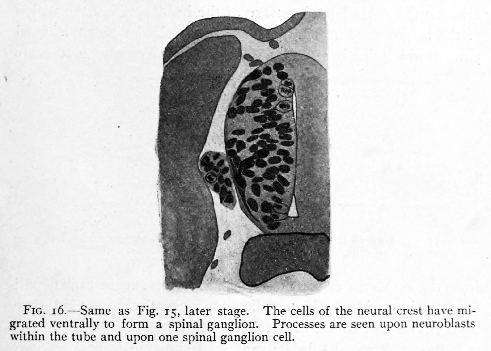 | 2,080 × 1,487 (331 KB) | Z8600021 (talk | contribs) |
You cannot overwrite this file.
File usage
The following 2 pages use this file: