File:Dextrocardia heart position.jpg
Dextrocardia_heart_position.jpg (400 × 533 pixels, file size: 49 KB, MIME type: image/jpeg)
Dextrocardia
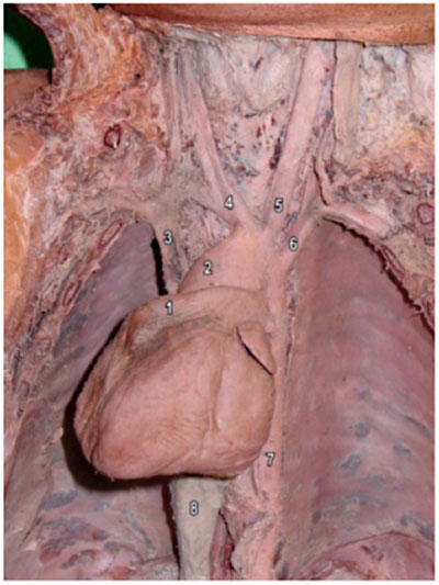
Anatomical position of the heart in the thorax.
During the inspection of the pulmonary trunk, a large ductus arteriosus (Botallo's ductus) was found. The heart apex was positioned along the heart axis and was turned to the right side, thus demonstrating the dextrocardia.
- 1 - pulmonary trunk
- 2 - ascending aorta
- 3 - superior vena cava (SVC)
- 4 - brachiocephalic trunk
- 5 - left common carotid artery
- 6 - left subclavial artery
- 7 - thoracic aorta
- 8 - hepatic tissue covering the inferior vena cava (IVC)
Original File Name: En_a13fig01.jpg female child, probably one year of age, which belonged to the Laboratory of Anatomy of the Campus of São José dos Campos - UNESP.
<pubmed>19142355</pubmed>| Arq Bras Cardiol.
All the content of the journal, except where otherwise noted, is licensed under a Creative Commons License.
File history
Yi efo/eka'e gwa ebo wo le nyangagi wuncin ye kamina wunga tinya nan
| Gwalagizhi | Nyangagi | Dimensions | User | Comment | |
|---|---|---|---|---|---|
| current | 00:48, 7 August 2010 | | 400 × 533 (49 KB) | S8600021 (talk | contribs) | ==Dextrocardia== Anatomical position of the heart in the thorax. During the inspection of the pulmonary trunk, a large ductus arteriosus (Botallo's ductus) was found. The heart apex was positioned along the heart axis and was turned to the right side, th |
You cannot overwrite this file.
File usage
The following 4 pages use this file: