File:Hassall1849 plate41.jpg
Original file (1,280 × 2,075 pixels, file size: 857 KB, MIME type: image/jpeg)
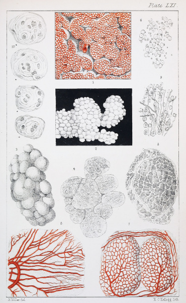
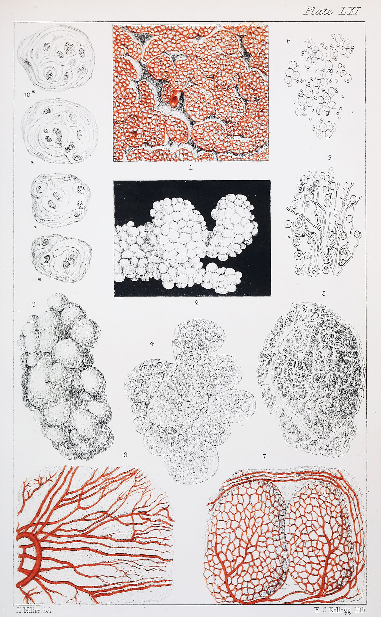
Plate LXI.
Fig. 1. Vessels of thyroid gland. 18 diameters.
Fig. 2. Vesicles of slightly enlarged thyroid, viewed with a lens only.
Fig. 3. Ditto of same, magnified 40 diameters.
Fig. 4. Ditto of same, magnified 67 diameters, showing the fibrous structure of their walls, and their cellular and nuclear contents.
Fig. 5. Lobes and vesicles of thyroid, magnified 27 diameters, as seen in a gland in its ordinary condition.
Fig. 6. Granular nuclei of vesicles of thyroid. Magnified 378 diameters.
Fig. 7. Two follicles of thymus gland, magnified 33 diameters, showing the plexus of vessels which invests them.
Fig. 8. A portion of the capsule of thymus, magnified 54 diameters, showing the ternary disposition of the vessels.
Fig. 9. Granular nuclei and simple cells with fibrous tissue of thymus. Magnified 378 diameters.
Fig. 10. Compound cells of thymus. Magnified 378 diameters.
Original scanned file name - Microscopicanato12hass_orig_0811.jp2
Reference
Hassall AH. The microscopic anatomy of the human body, in health and disease. (1849) Samuel Hurley, Fleet Street, London.
File history
Yi efo/eka'e gwa ebo wo le nyangagi wuncin ye kamina wunga tinya nan
| Gwalagizhi | Nyangagi | Dimensions | User | Comment | |
|---|---|---|---|---|---|
| current | 12:29, 22 Ocak 2019 | | 1,280 × 2,075 (857 KB) | Z8600021 (talk | contribs) | |
| 12:27, 22 Ocak 2019 | 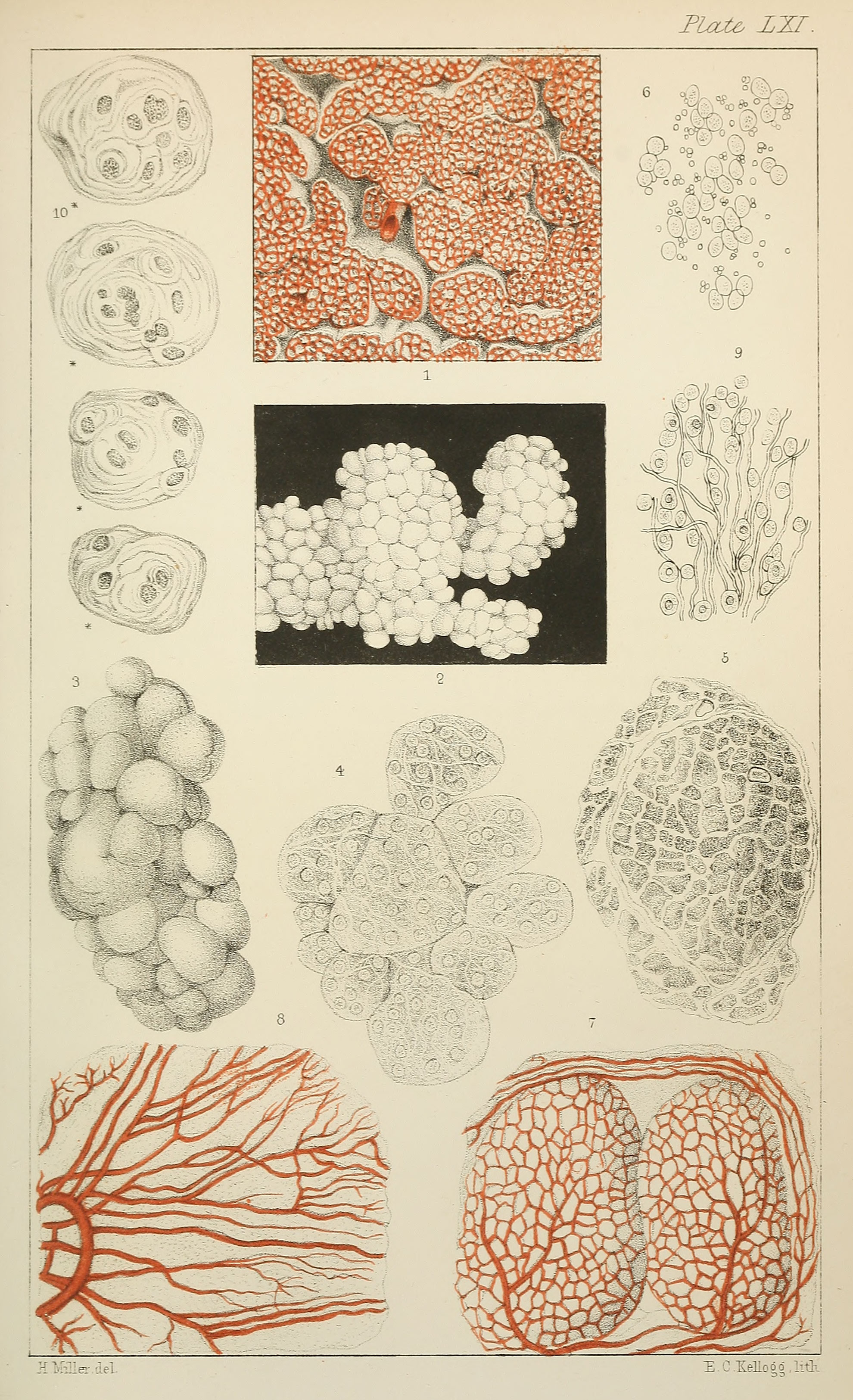 | 1,980 × 3,250 (1.28 MB) | Z8600021 (talk | contribs) | ==Plate LXI. == Original scanned file name - Microscopicanato12hass_orig_0811.jp2 ===Reference=== {{Ref-Hassall1849 }} |
You cannot overwrite this file.
File usage
The following page uses this file: