File:Cullen1916 fig15.jpg
Original file (1,000 × 1,227 pixels, file size: 314 KB, MIME type: image/jpeg)
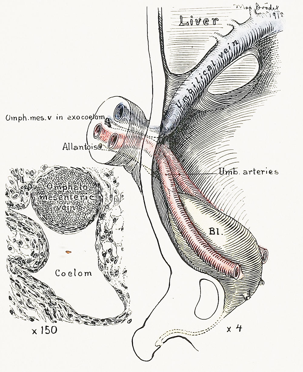
Fig. 15. Sagittal View of a Graphic Reconstruction of the Umbilical Region of a Human Embryo 5.2 cm in Length
From within, the relations of the umbilical arteries to the vein are clearly seen. They surround the funnel-shaped remnant of the exoccelom. No trace of the omphalomesenteric vessels is to be seen in the peritoneal cavity, but in cross-sections of the cord near the embryo the lumen of the umbilical vein still persists, as indicated in the small drawing to the left. It is embedded in the wall of the exoccelom, but has no connection with any other vascular structure. The bladder bulges out between the umbilical arteries. The urachus (allantois) passes out into the cord between and below the umbilical arteries. This is the characteristic arrangement. The urachus shows two dilatations.
File history
Yi efo/eka'e gwa ebo wo le nyangagi wuncin ye kamina wunga tinya nan
| Gwalagizhi | Nyangagi | Dimensions | User | Comment | |
|---|---|---|---|---|---|
| current | 13:13, 28 October 2018 | | 1,000 × 1,227 (314 KB) | Z8600021 (talk | contribs) | |
| 13:12, 28 October 2018 |  | 2,091 × 1,714 (676 KB) | Z8600021 (talk | contribs) | Fig. 15. Sagittal View of a Graphic Reconstruction of the Umbilical Region of a Human Embryo 5.2 cm. in Length. From within, the relations of the umbilical arteries to the vein are clearly seen. They surround the funnel-shaped remnant of the exoccel... |
You cannot overwrite this file.
File usage
The following 2 pages use this file: