File:Bandler1983 fig05.jpg
From Embryology
Size of this preview: 609 × 599 pixels. Other resolution: 1,000 × 984 pixels.
Original file (1,000 × 984 pixels, file size: 236 KB, MIME type: image/jpeg)
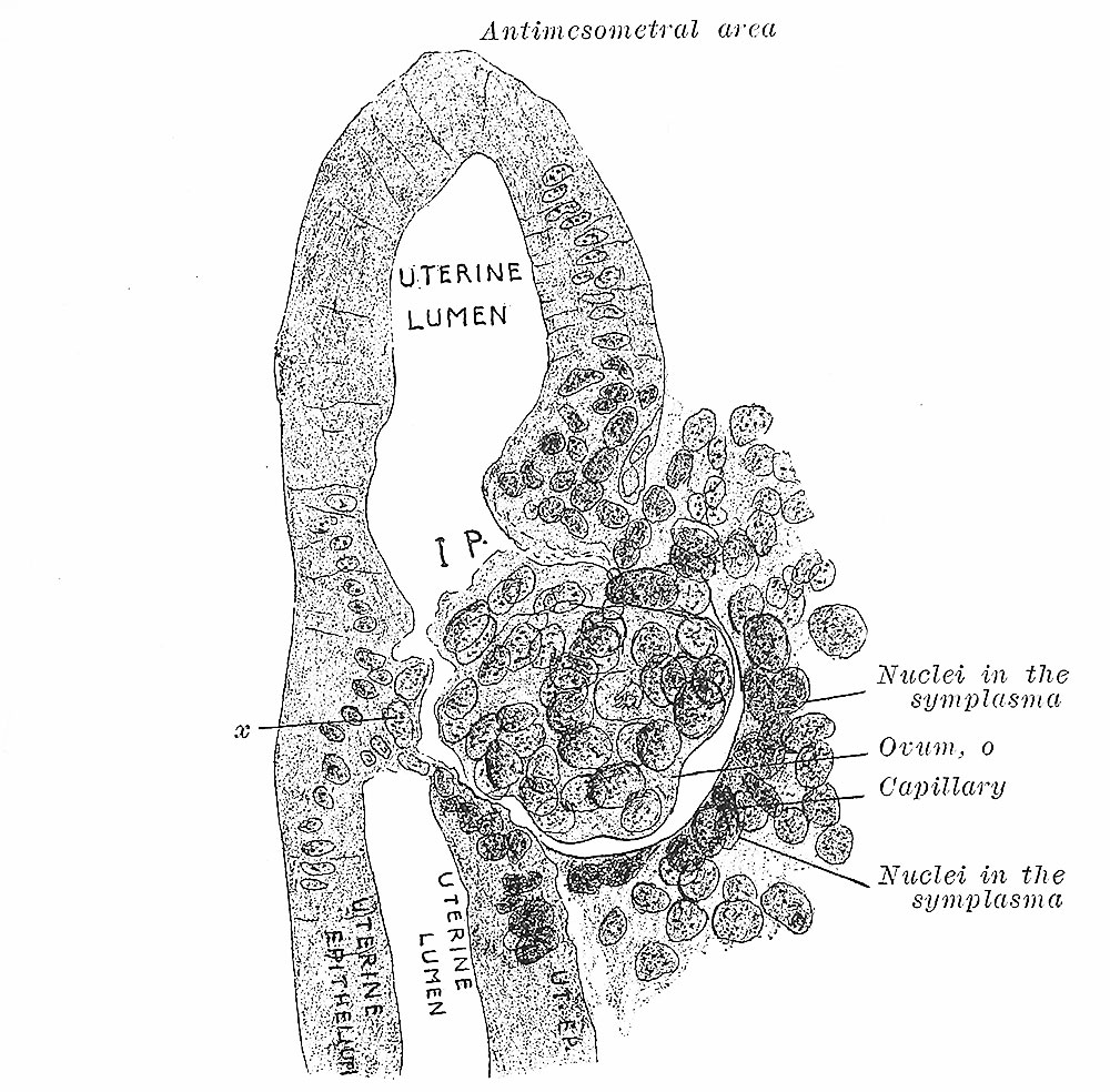
Fig. 5. Further stage in embedding of ovum
Showing changes wrought in the connective tissue compacta. (v. Spee.)
x, two cells of the ovum which are embedded on the opposite side.
Reference
Bandler SW. Uterine and Tubal Gestation (1893) William Wood & Company, New York.
Cite this page: Hill, M.A. (2024, June 26) Embryology Bandler1983 fig05.jpg. Retrieved from https://embryology.med.unsw.edu.au/embryology/index.php/File:Bandler1983_fig05.jpg
- © Dr Mark Hill 2024, UNSW Embryology ISBN: 978 0 7334 2609 4 - UNSW CRICOS Provider Code No. 00098G
File history
Yi efo/eka'e gwa ebo wo le nyangagi wuncin ye kamina wunga tinya nan
| Gwalagizhi | Nyangagi | Dimensions | User | Comment | |
|---|---|---|---|---|---|
| current | 22:58, 11 September 2018 | | 1,000 × 984 (236 KB) | Z8600021 (talk | contribs) | |
| 22:56, 11 September 2018 | 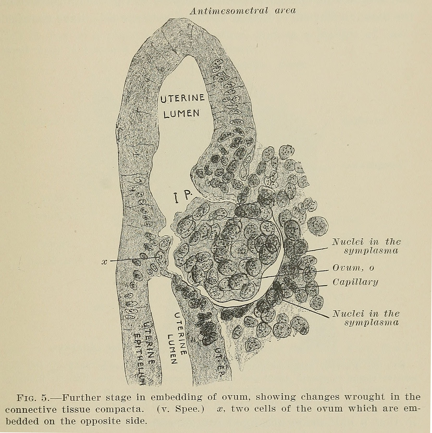 | 1,658 × 1,663 (536 KB) | Z8600021 (talk | contribs) | ===Reference=== {{Ref-Bandler1983}} {{Footer}} |
You cannot overwrite this file.
File usage
The following page uses this file: