File:Locy1895 plate27.jpg

Original file (4,342 × 2,850 pixels, file size: 477 KB, MIME type: image/jpeg)
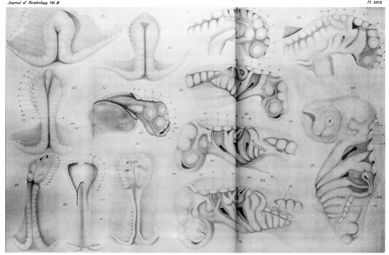
Plate XXVII
All the figures are of Squalus acanthia they are drawn with the aid of thecamera, and are all d( 45 diameters.
Fig. 25. Young embryo intermediate between Balfoufs stages B and C. The embryo so far as formed is divided into eight pairs of metameres and these arecontinued without brealc, or any change in Character, into the halves of the embryonic rim. The eleventh Inetamere which, in later stages, lies in kront of the vagus nerve is now on either side the third one from the axial embryo.
Fig. 26. Somewhat older embryo, showing the change in form of the head region. The axial embryo now includes about fifteen pairs of rnetamerea
Fig. 27. Slightly older than the precedingz there are about eighteen pairs of rnetameres in the axial part of the embryo and,·as in the forrner instances, are continued into the embryonic rim. Two longitudinal marginal furrows have appeared, separating two marginal bands from the rest of the embryo. Along the line of these furrows are seen four depressions that mark the very beginning of segmental Sense-Organs.
FIG. 28. View from the upper side of the same embryo illustrated in Fig. 29. The cephalic plate is now clearly marked off from the more slender trunk region. The depressions for the optic vesicles (c-P.) have made their appearancqz
F1G. 29. View of the same embryo from the ventral aspect. The yolk has been cornpletely removed, and we get a view directly into the gastrular cavity. There are eleven pairs of metameres in the broad part of the cephalic plate. The neural folds are ventrally curved The outlines of the figure and the neural segments are too symmetrical.
FIG. 30. Older embryo with neural folds lying in the horizontal plane. The broad cephalic plate is in marked contrast with the slender trank.
Fig. 31. Embryo in which the neural folds have nearly attained the vertical Plane. The neural groove is still open. The optic vesicle (qk).) and the combined vesicle of mid—brain and accessory optic Eis-z. -l- A. tax-L) vesicle show on the sides of the head. There is also the beginning of rnandibnlar head cavity El. c) and the branchia1 pouch. The original Inetameric divisions are still very plain.
FIG. 32. Embryo just after the closure of the neural groove in the anterior end. The posterior part of the neural canal is not cornpletely closed. Note the metarneric divisions indicated by numbers J, e, F, etc. ·
Fig. 33. Embryo after complete closure of the neural groove and before the appearance of the ear vesicle.
Fig. 34. Embryo after the differentiation of the ear saucen The five anterior metarneric divisions are no longer distinguishable, those of the hind-brain are prominent and are approximatecl in the middle plane. 0ne gilbcleft has broken through. The nasal pit has started. ·
Fig. 35. slightly older embryo showing several characteristic changea The— line of neural segments are being forced apart by lateralgrowth of the roof of the hind-brain. The fifth nerve is plainly visib1e. Over the gillsclefts runs a continuous nodulated thiclcening containing the branchial Sense-Organs and the radirnent of the lateral 1ine. 588 Lock:
Fig. 36. Some-what older embryo differing from the preceding mainly in show ing the rudiments of the seventh, eighth ninth, and tenth next-es. Note the lens and choroid Hssure in the eye vesic1e.
Fig. 37. Slightly older than the precedjng The line of neural segments are undergoing some ehanges whereby the concavity on the lower margin is made to correspond vvith a crest on the upper margitx In embryos of about the age represented in Fig. 36 or a very little older the epiphysial outgrowth arises from the roof of the tha1amencepha1on.
- Locy 1895 Contents: General Introduction | Part I - Metamerism of the Head | Part II - The Sense-Organs | Figures
Reference
Locy WA.Contribution to the structure and development of the vertebrate head. (1895) J. Morphol. 11(3): 497-595.
Cite this page: Hill, M.A. (2024, June 24) Embryology Locy1895 plate27.jpg. Retrieved from https://embryology.med.unsw.edu.au/embryology/index.php/File:Locy1895_plate27.jpg
- © Dr Mark Hill 2024, UNSW Embryology ISBN: 978 0 7334 2609 4 - UNSW CRICOS Provider Code No. 00098G
File history
Yi efo/eka'e gwa ebo wo le nyangagi wuncin ye kamina wunga tinya nan
| Gwalagizhi | Nyangagi | Dimensions | User | Comment | |
|---|---|---|---|---|---|
| current | 15:43, 27 August 2018 | | 4,342 × 2,850 (477 KB) | Z8600021 (talk | contribs) | |
| 15:42, 27 August 2018 |  | 4,342 × 2,850 (354 KB) | Z8600021 (talk | contribs) |
You cannot overwrite this file.
File usage
The following page uses this file: