File:LewisFT1920 fig10-13.jpg
Original file (2,348 × 3,299 pixels, file size: 442 KB, MIME type: image/jpeg)
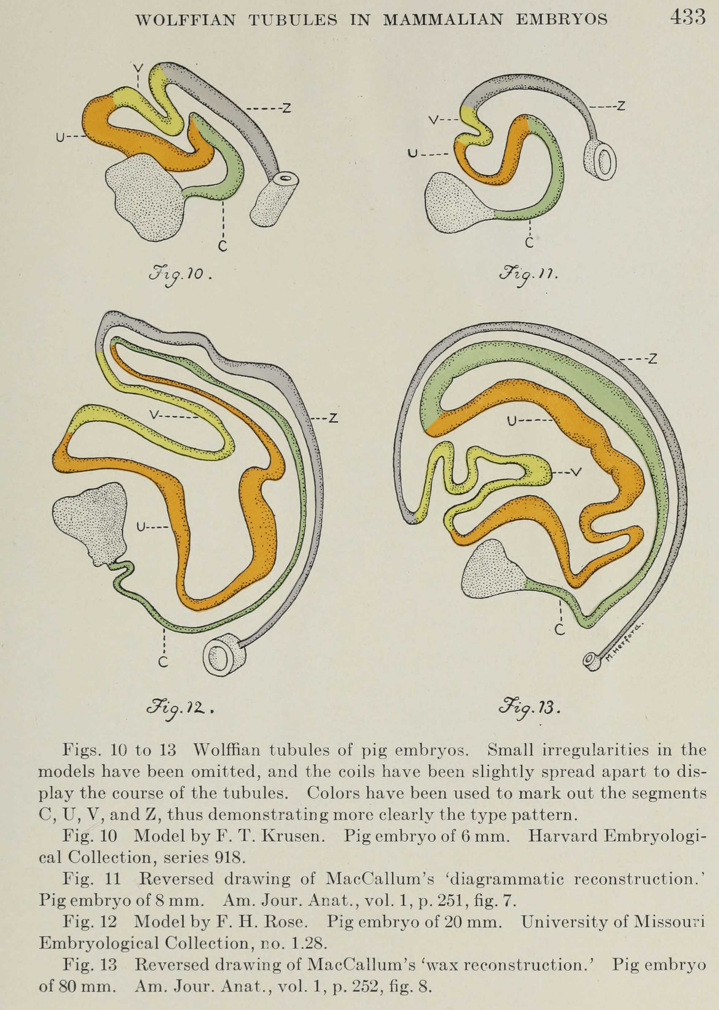
Fig. 10 Model by F. T. Krusen. Pig embryo of 6 mm. Harvard Embryologi- cal Collection, series 918.
Fig. 11 Reversed drawing of MacCallum’s ‘diagrammatic reconstruction. Pig embryo of 8 mm. Am. Jour. Anat., vol. 1, p. 251, fig. 7.
Fig. 12 Model by F. H. Rose. Pig embryo of 20 mm. University of Missouri Embryological Collection, no. 1.28.
Fig. 13 Reversed drawing of MacCallum’s ‘wax reconstruction. Pig embryo of 80mm. Am. Jour. Anat., vol. 1, p. 252, fig. 8.
Reference
Lewis FT. The course of the Wolffian tubules in mammalian embryos. (1920) Amer. J Anat. 26(3): 423-436.
Cite this page: Hill, M.A. (2026, October 4) Embryology LewisFT1920 fig10-13.jpg. Retrieved from https://embryology.med.unsw.edu.au/embryology/index.php/File:LewisFT1920_fig10-13.jpg
- © Dr Mark Hill 2026, UNSW Embryology ISBN: 978 0 7334 2609 4 - UNSW CRICOS Provider Code No. 00098G
File history
Yi efo/eka'e gwa ebo wo le nyangagi wuncin ye kamina wunga tinya nan
| Gwalagizhi | Nyangagi | Dimensions | User | Comment | |
|---|---|---|---|---|---|
| current | 15:32, 9 August 2018 | | 2,348 × 3,299 (442 KB) | Z8600021 (talk | contribs) | ===Reference=== {{Ref-LewisFT1920}} {{Footer}} |
You cannot overwrite this file.
File usage
The following page uses this file:


