File:Fetal gonad retinoid receptor expression 01.jpg
Original file (1,004 × 1,000 pixels, file size: 226 KB, MIME type: image/jpeg)
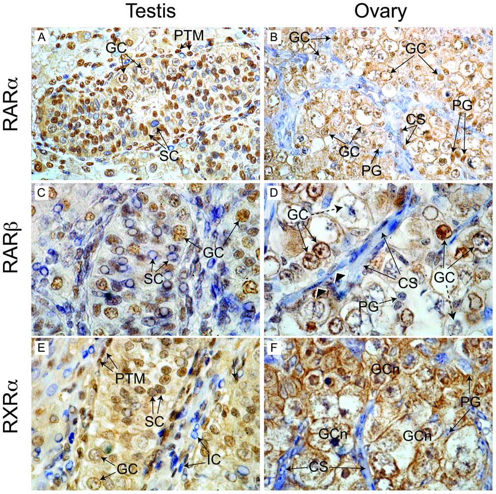
Human Fetal Gonad Retinoid Receptor Expression
Second trimester human fetal testis and ovary.
|
|
|
|
The widespread nuclear localization of RA receptors in testis suggests cells of all types (including germ cells) are exposed to RA signals. |
|
Magnification: 400× (A, B), 1000× (C–F).
Reference
<pubmed>21674038</pubmed>
Copyright: © 2011 Childs et al. This is an open-access article distributed under the terms of the Creative Commons Attribution License, which permits unrestricted use, distribution, and reproduction in any medium, provided the original author and source are credited.
Text edited from original figure legend. Figure 3. doi:info:doi/10.1371/journal.pone.0020249.g003
File history
Yi efo/eka'e gwa ebo wo le nyangagi wuncin ye kamina wunga tinya nan
| Gwalagizhi | Nyangagi | Dimensions | User | Comment | |
|---|---|---|---|---|---|
| current | 17:48, 21 May 2012 | | 1,004 × 1,000 (226 KB) | Z8600021 (talk | contribs) | Immunohistochemical localisation of retinoid receptor expression in the human fetal gonad. In the second trimester human fetal testis (A) RARα staining was detected in germ cell (GC) and peritubular myoid (PTM) nuclei. Two populations of Sertoli cells ( |
You cannot overwrite this file.
File usage
The following 2 pages use this file: