File:Fawcett1975 fig31.jpg
From Embryology

Size of this preview: 800 × 252 pixels. Other resolution: 1,280 × 403 pixels.
Original file (1,280 × 403 pixels, file size: 128 KB, MIME type: image/jpeg)
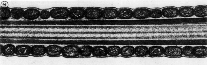
FIG. 31. A longitudinal thin section of the middle piece of a mammalian spermatozoon. The circumferen- tially oriented mitochondria are cut transversely. Note how closely the celi membrane is apposed to the underlying mitochondria.
File history
Yi efo/eka'e gwa ebo wo le nyangagi wuncin ye kamina wunga tinya nan
| Gwalagizhi | Nyangagi | Dimensions | User | Comment | |
|---|---|---|---|---|---|
| current | 11:02, 20 August 2017 | | 1,280 × 403 (128 KB) | Z8600021 (talk | contribs) | |
| 11:02, 20 August 2017 | 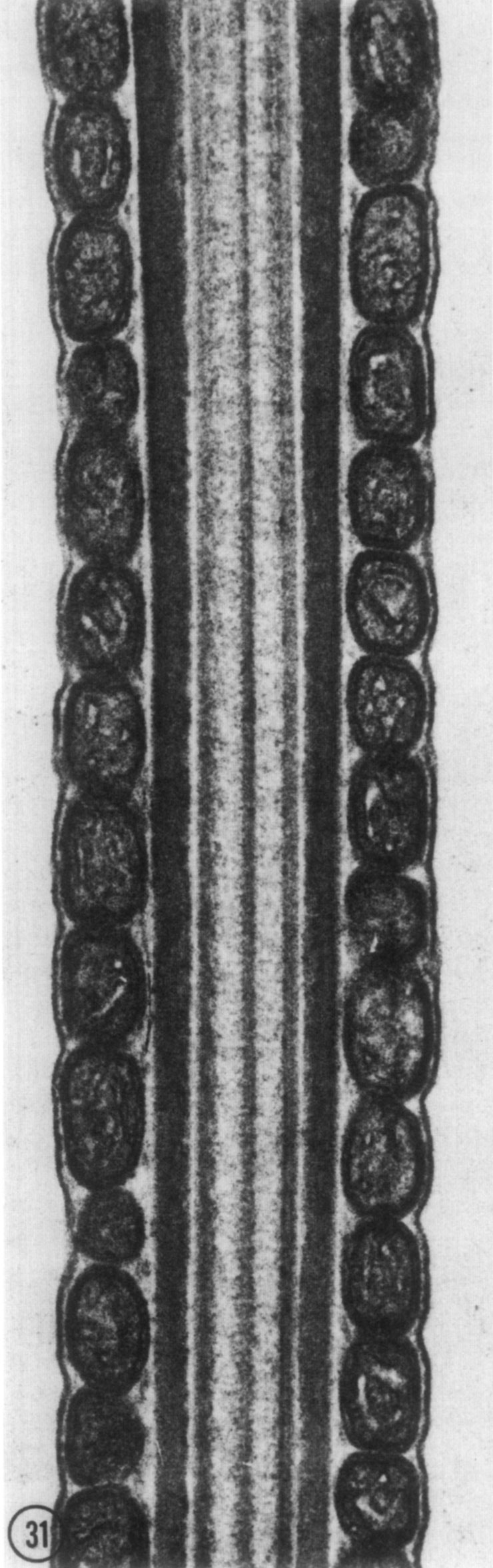 | 740 × 2,353 (269 KB) | Z8600021 (talk | contribs) | FIG. 31. A longitudinal thin section of the middle piece of a mammalian spermatozoon. The circumferen- tially oriented mitochondria are cut transversely. Note how closely the celi membrane is apposed to the underlying mitochondria. |
You cannot overwrite this file.
File usage
The following page uses this file: