File:Cameron1916 fig01.jpg
Original file (586 × 1,000 pixels, file size: 108 KB, MIME type: image/jpeg)
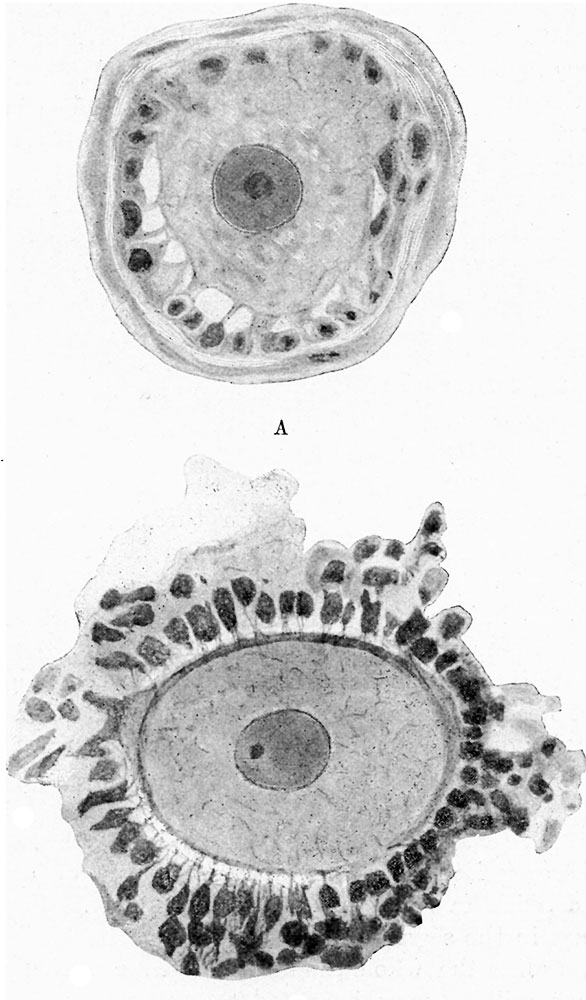
Fig. 1. Two stages illustrating the development of the ovarian ovum and Graafian follicle in the cat
A shows an early stage, before the complete development of the zona pellucida. It will be observed that the c toplasm of the follicle cells is. continuous, not only with that of the ovum, but a so with that of the adjacent cells of the same layer, and of the ovarian stroma. B, later stage, in which it will be noted that the zona pellucida now forms a distinct boundary zone, but does not completely separate the ovum from the cells of the corona radiate. Processes from those cells pass through the zona pellucida, and are continuous with theoyto-reticulum of the ooplasm.
Reference
Cameron J. and Gladstone RJ. The structure of the blastoderm, and the continuity of the cell-elements during the early stages of development. (1916) J Anat. Physiol. 50(3): 207-27. PubMed 17233062
Cite this page: Hill, M.A. (2024, June 26) Embryology Cameron1916 fig01.jpg. Retrieved from https://embryology.med.unsw.edu.au/embryology/index.php/File:Cameron1916_fig01.jpg
- © Dr Mark Hill 2024, UNSW Embryology ISBN: 978 0 7334 2609 4 - UNSW CRICOS Provider Code No. 00098G
File history
Yi efo/eka'e gwa ebo wo le nyangagi wuncin ye kamina wunga tinya nan
| Gwalagizhi | Nyangagi | Dimensions | User | Comment | |
|---|---|---|---|---|---|
| current | 11:43, 12 August 2017 | | 586 × 1,000 (108 KB) | Z8600021 (talk | contribs) | |
| 11:42, 12 August 2017 |  | 1,315 × 1,953 (337 KB) | Z8600021 (talk | contribs) |
You cannot overwrite this file.
File usage
The following page uses this file: