File:Boyd1950 fig03a.jpg
From Embryology
Size of this preview: 412 × 599 pixels. Other resolution: 600 × 872 pixels.
Original file (600 × 872 pixels, file size: 135 KB, MIME type: image/jpeg)
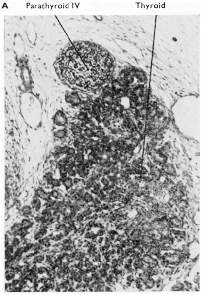
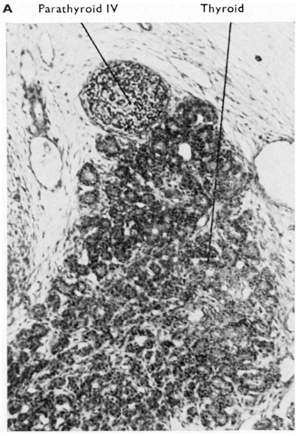
Fig. 3A. Transverse section through thyroid and parathyroid IV of a 60 mm human embryo
(x 350.) The thyroid tissue shows developing follicles and commencing appearance of colloid in them.
Reference
Boyd JD. Development of the thyroid and parathyroid glands and the thymus. (1950) Ann R Coll Surg Engl. 7(6): 455-71. PMID 14790564
Cite this page: Hill, M.A. (2026, October 4) Embryology Boyd1950 fig03a.jpg. Retrieved from https://embryology.med.unsw.edu.au/embryology/index.php/File:Boyd1950_fig03a.jpg
- © Dr Mark Hill 2026, UNSW Embryology ISBN: 978 0 7334 2609 4 - UNSW CRICOS Provider Code No. 00098G
File history
Yi efo/eka'e gwa ebo wo le nyangagi wuncin ye kamina wunga tinya nan
| Gwalagizhi | Nyangagi | Dimensions | User | Comment | |
|---|---|---|---|---|---|
| current | 23:43, 6 March 2017 | | 600 × 872 (135 KB) | Z8600021 (talk | contribs) | |
| 23:42, 6 March 2017 | 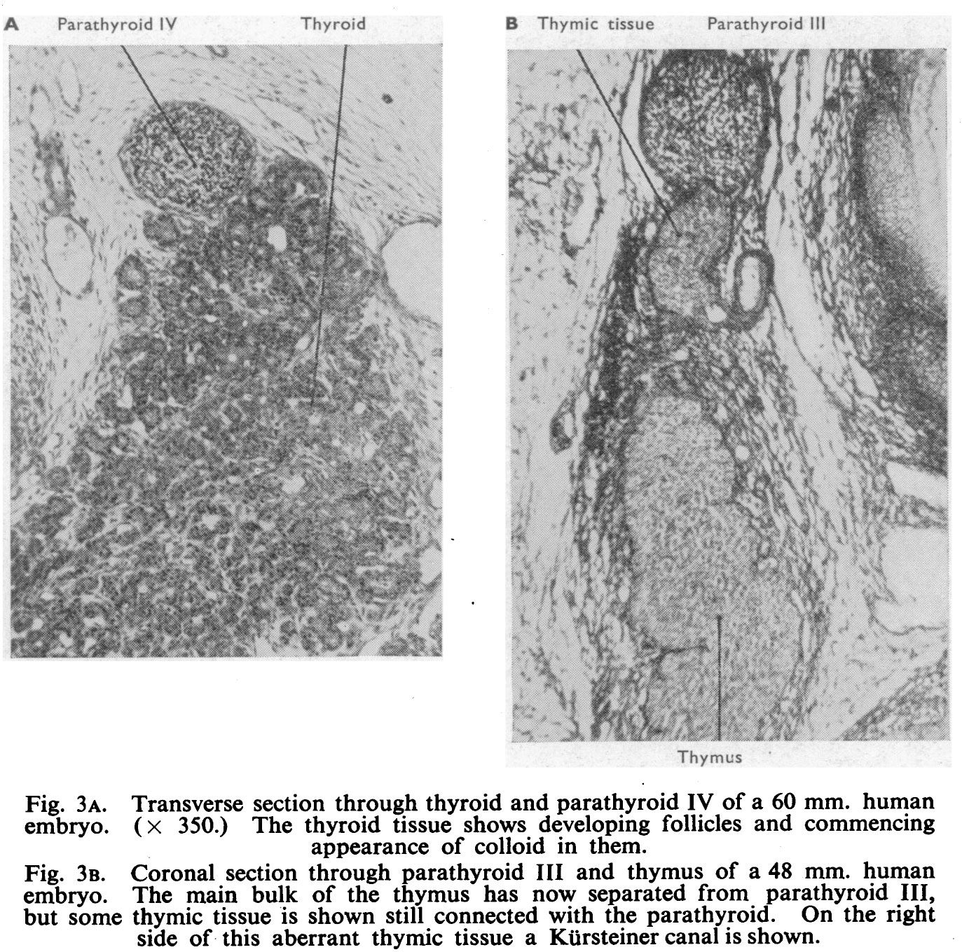 | 1,373 × 1,359 (566 KB) | Z8600021 (talk | contribs) |
You cannot overwrite this file.
File usage
The following page uses this file: