File:Frazer1911 fig03.jpg
From Embryology

Size of this preview: 800 × 375 pixels.
Original file (1,280 × 600 pixels, file size: 153 KB, MIME type: image/jpeg)
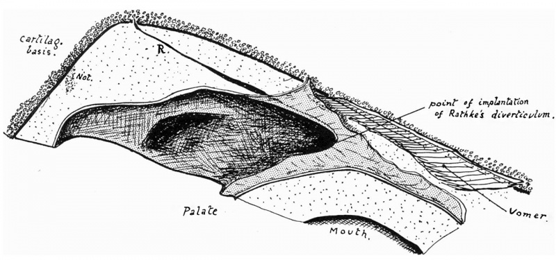
Fig. 3. The mucous membrane right nasal opening 4 months
The mucous membrane of the right nasal opening is shown cut, marking the upper limxt of the opening. Traced from a linear reconstruction. x 18. 4 months.
Reference
Frazer JE. Pharyngeal end of Rathke's pouch. (1911) J Anat. 45: 190-196. PMID 17232879
Cite this page: Hill, M.A. (2026, October 3) Embryology Frazer1911 fig03.jpg. Retrieved from https://embryology.med.unsw.edu.au/embryology/index.php/File:Frazer1911_fig03.jpg
- © Dr Mark Hill 2026, UNSW Embryology ISBN: 978 0 7334 2609 4 - UNSW CRICOS Provider Code No. 00098G
File history
Yi efo/eka'e gwa ebo wo le nyangagi wuncin ye kamina wunga tinya nan
| Gwalagizhi | Nyangagi | Dimensions | User | Comment | |
|---|---|---|---|---|---|
| current | 11:15, 9 Ocak 2017 | | 1,280 × 600 (153 KB) | Z8600021 (talk | contribs) | |
| 11:15, 9 Ocak 2017 |  | 1,530 × 817 (215 KB) | Z8600021 (talk | contribs) | ===Reference=== {{Ref-Frazer1911}} {{Footer}} |
You cannot overwrite this file.
File usage
The following 2 pages use this file: