File:Frazer1911 fig02.jpg
Original file (1,280 × 872 pixels, file size: 112 KB, MIME type: image/jpeg)
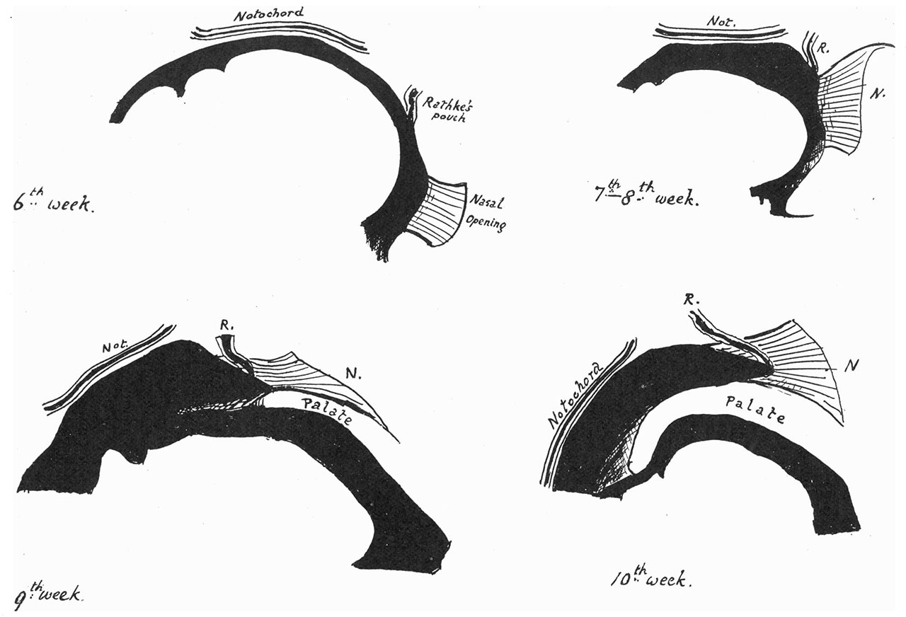
Fig. 2 Pharyngeal end of Rathke's pouch
The diagrams in fig. 2 are drawn to scale from models, and are intended to illustrate the position of the pouch and notochord in a median sagittal section of each embryo, the anterior end being to the right. The situation of the internal nasal openings is also indicated, and it can be seen that these tend rapidly to assume a higher position, until they come to open on a level with the roof of the pharynx.
Reference
Frazer JE. Pharyngeal end of Rathke's pouch. (1911) J Anat. 45: 190-196. PMID 17232879
Cite this page: Hill, M.A. (2026, October 5) Embryology Frazer1911 fig02.jpg. Retrieved from https://embryology.med.unsw.edu.au/embryology/index.php/File:Frazer1911_fig02.jpg
- © Dr Mark Hill 2026, UNSW Embryology ISBN: 978 0 7334 2609 4 - UNSW CRICOS Provider Code No. 00098G
File history
Yi efo/eka'e gwa ebo wo le nyangagi wuncin ye kamina wunga tinya nan
| Gwalagizhi | Nyangagi | Dimensions | User | Comment | |
|---|---|---|---|---|---|
| current | 11:07, 9 Ocak 2017 | | 1,280 × 872 (112 KB) | Z8600021 (talk | contribs) | |
| 11:07, 9 Ocak 2017 | 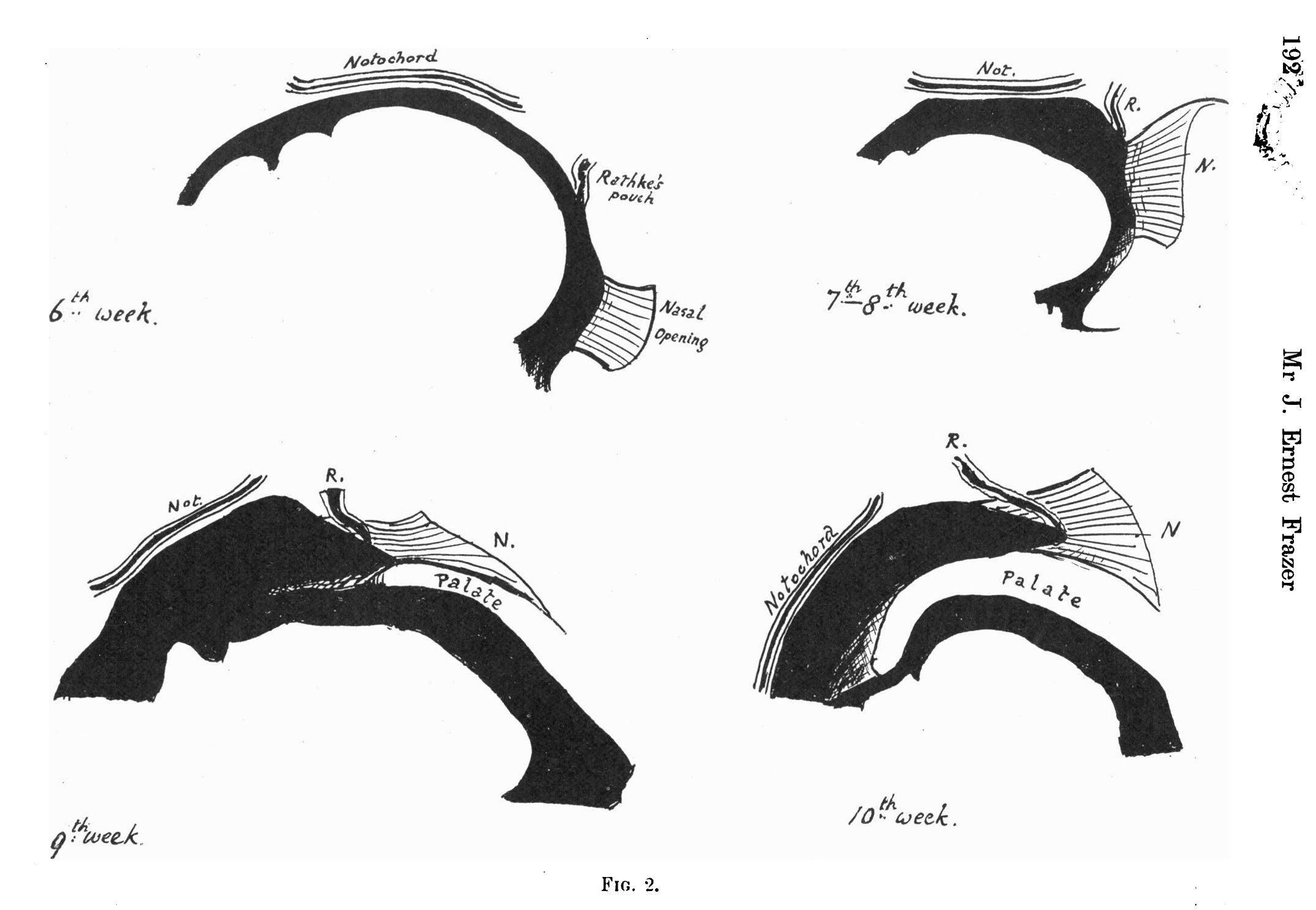 | 2,152 × 1,488 (226 KB) | Z8600021 (talk | contribs) | ===Reference=== {{Ref-Frazer1911}} {{Footer}} |
You cannot overwrite this file.
File usage
The following 2 pages use this file:


