File:Hunter1935 plate01.jpg
Original file (1,790 × 2,107 pixels, file size: 711 KB, MIME type: image/jpeg)
Explanation of Plate I
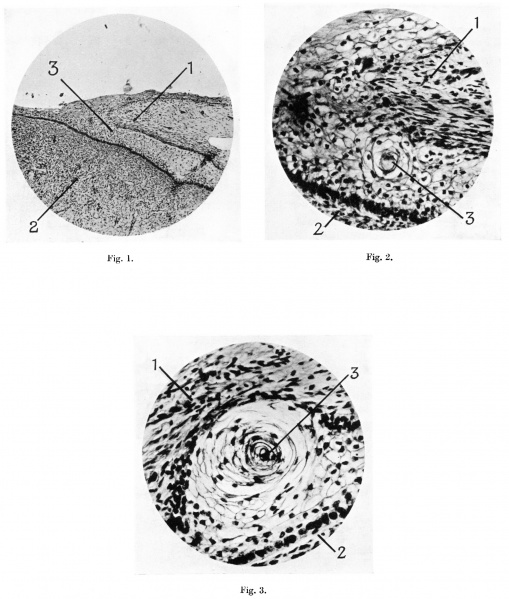
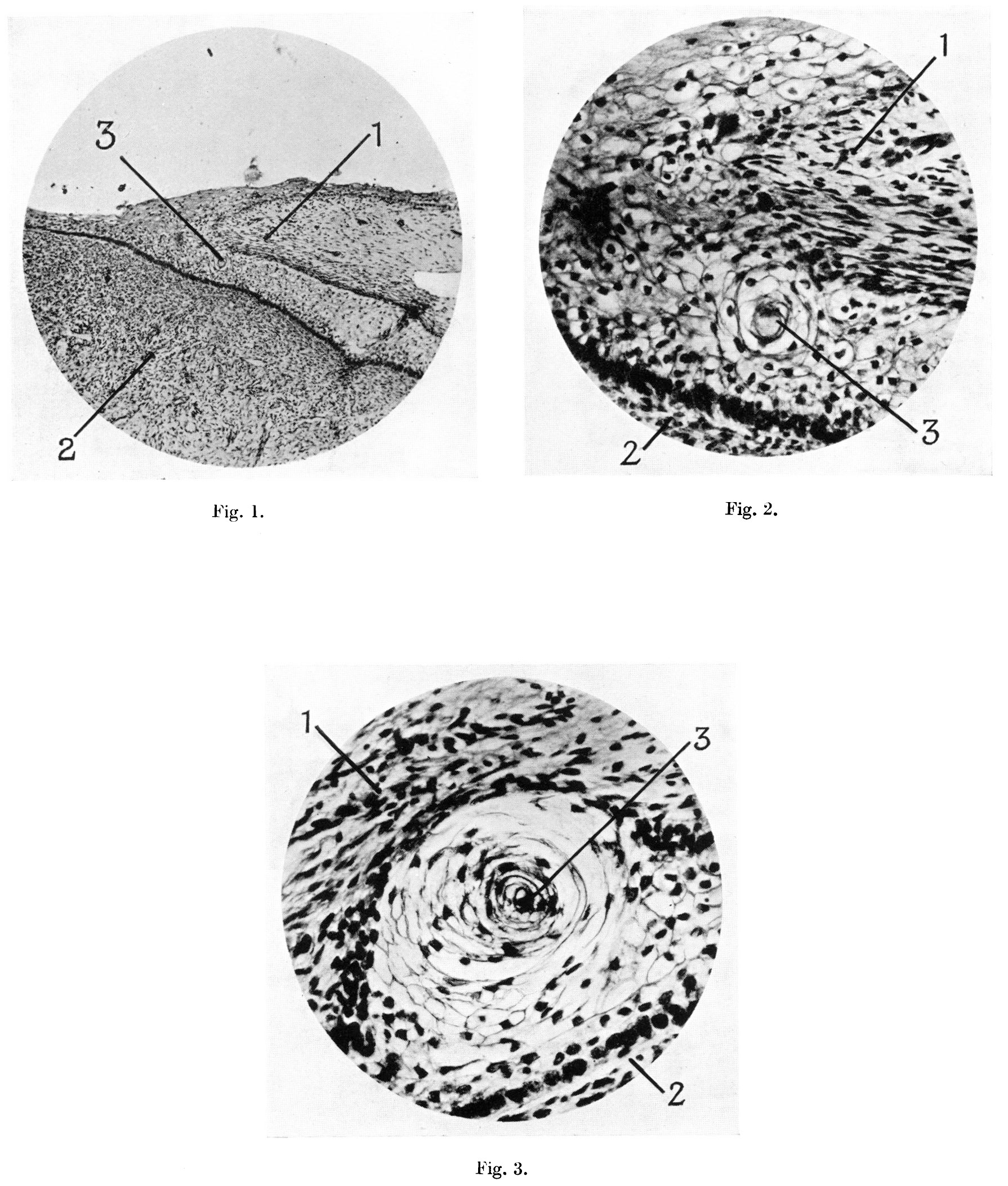
Fig. 1. Microphotograph of terminal part of penis from human foetus of 100 mm. C.R. length, to show the continuity of the cells of the epitriehial layer of the prepuee with those of the layer between the prepuee and glans. Pointers: 1, prepuce; 2, glans penis; 3, cell nest.
Fig. 2. Microphotograph of a “cell-nest” in the layer of cells between prepuce and glans, from a human foetus of 100 mm. C.R. length. Pointers as fig. 1.
Fig. 3. Microphotograph of a “cell-nest” in the layer of cell between prepuce and glans, from a human foetus of 170 mm. C.R.. length. Pointers as fig. 1.
File history
Yi efo/eka'e gwa ebo wo le nyangagi wuncin ye kamina wunga tinya nan
| Gwalagizhi | Nyangagi | Dimensions | User | Comment | |
|---|---|---|---|---|---|
| current | 17:27, 4 Ocak 2017 | | 1,790 × 2,107 (711 KB) | Z8600021 (talk | contribs) | |
| 17:26, 4 Ocak 2017 | 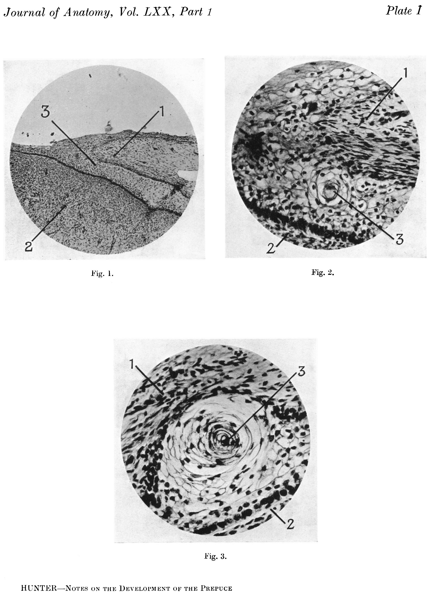 | 1,790 × 2,452 (802 KB) | Z8600021 (talk | contribs) | ==Explanation of Plate I== Fig. 1. Microphotograph of terminal part of penis from human foetus of 100 mm. C.R. length, to show the continuity of the cells of the epitriehial layer of the prepuee with those of the layer between the prepuee and glans. P... |
You cannot overwrite this file.
File usage
The following page uses this file: