File:Cooper1938 plate30.jpg

Original file (1,655 × 2,457 pixels, file size: 402 KB, MIME type: image/jpeg)
Plate XXXI
Fig. 17. Swollen cells lining an alveolus in the bronchopneumonic lung of an infant aged 12 weeks. X 630.
Fig. 18. Cells lining alveoli in the lung of an infant aged 8 months. R.B.C. = erythrocyte. X700.
Fig. 19. Lung of a three-days-old mouse showing continuous lining of cells in the alveoli. R.B.C. = erythrocyte. X625.
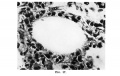
Fig. 17. infant aged 12 weeks
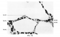
Fig. 18. infant aged 8 months
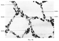
Fig. 19. three-days-old mouse


| Historic Disclaimer - information about historic embryology pages |
|---|
 |
Reference
Cooper ERA. A histological investigation of the development and structure of the human lung. (1938) J Pathology 47: 105-114.
Cite this page: Hill, M.A. (2024, June 21) Embryology Cooper1938 plate30.jpg. Retrieved from https://embryology.med.unsw.edu.au/embryology/index.php/File:Cooper1938_plate30.jpg
- © Dr Mark Hill 2024, UNSW Embryology ISBN: 978 0 7334 2609 4 - UNSW CRICOS Provider Code No. 00098G
File history
Yi efo/eka'e gwa ebo wo le nyangagi wuncin ye kamina wunga tinya nan
| Gwalagizhi | Nyangagi | Dimensions | User | Comment | |
|---|---|---|---|---|---|
| current | 10:12, 27 November 2016 | | 1,655 × 2,457 (402 KB) | Z8600021 (talk | contribs) | ==Plate XXXI== Fig. 17. Swollen cells lining an alveolus in the bronchopneumonic lung of an infant aged 12 weeks. X 630. Fig. 18. Cells lining alveoli in the lung of an infant aged 8 month... |
You cannot overwrite this file.
File usage
The following page uses this file: