File:Human week 10 fetus 03.jpg
From Embryology
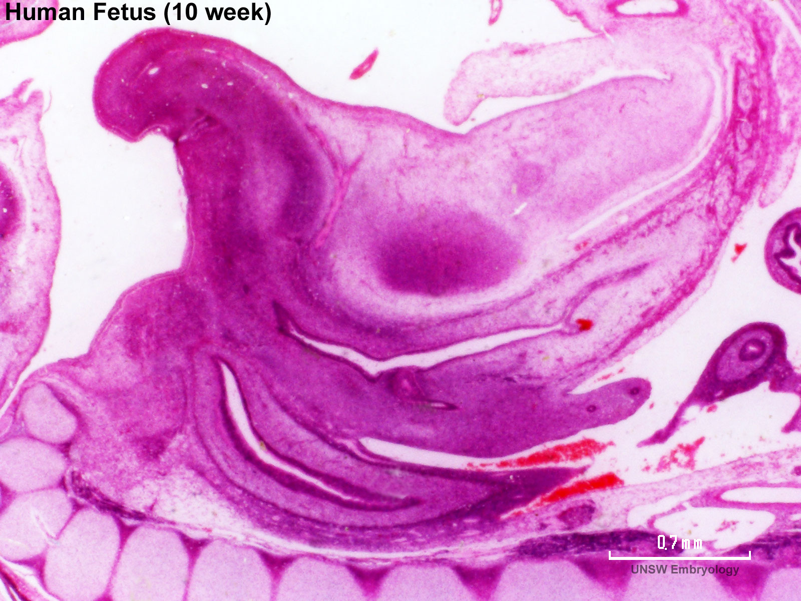
Size of this preview: 800 × 600 pixels. Other resolution: 1,600 × 1,200 pixels.
Original file (1,600 × 1,200 pixels, file size: 370 KB, MIME type: image/jpeg)
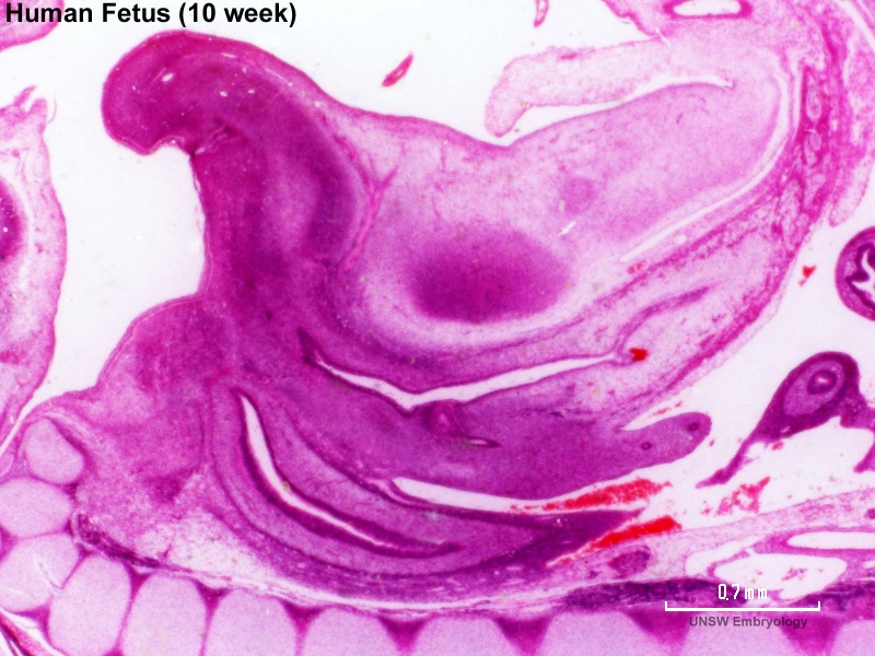
Human Female Fetus Pelvic Region (10 week)
Large image version of plane D, close to midline (Stain - Haematoxylin Eosin) 3 mm scale bar.
| Features |
|---|
|
- Human Female Fetus (week 10)
Sagittal Section (plane D)
Pituitary and Lamina Terminalis
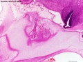
Olfactory Nerve
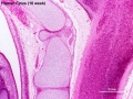
Atlas and Axis
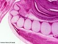
Sacrum
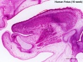
Oral Cavity
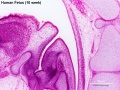
Epiglottis
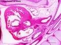
Heart
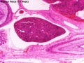
Spleen
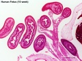
Midgut Herniation
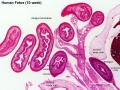
Midgut Herniation (label)
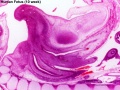
Pelvic Region
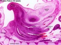
Pelvic Region (label)


Related Images

Fetus (week 10) Planes A (most lateral), B (lateral), C (medial) and D (midline) from lateral towards the midline.
- Human Fetus - most lateral | lateral | medial | midline
- Head - most lateral | lateral | medial | midline
- Cerebellum - most lateral | lateral | medial | midline
- Urogenital Unlabelled - most lateral | lateral | medial | midline
- Urogenital Labelled - most lateral | lateral | medial | midline
- Large Images - midline
- Image Source: UNSW Embryology, no reproduction without permission.
File history
Yi efo/eka'e gwa ebo wo le nyangagi wuncin ye kamina wunga tinya nan
| Gwalagizhi | Nyangagi | Dimensions | User | Comment | |
|---|---|---|---|---|---|
| current | 17:22, 17 Haziran 2012 | | 1,600 × 1,200 (370 KB) | Z8600021 (talk | contribs) | |
| 17:19, 17 Haziran 2012 |  | 1,200 × 900 (248 KB) | Z8600021 (talk | contribs) | ==Human Female Fetus Pelvic Region (10 week)== Large image version of plane D, close to midline (H&E stain). 3 mm scale bar {{10wkFetus}} |
You cannot overwrite this file.
File usage
The following 22 pages use this file:
- BGDA Practical 12 - Embryo to Fetus
- BGDB Gastrointestinal - Fetal
- BGDB Sexual Differentiation - Fetal
- Fetal Development - 10 Weeks
- Foundations Practical - Week 9 to 36
- G
- REI - Reproductive Medicine Seminar 2018
- Royal Hospital for Women - Reproductive Medicine Seminar 2018
- File:Human week 10 fetus 01.jpg
- File:Human week 10 fetus 03.jpg
- File:Human week 10 fetus 04.jpg
- File:Human week 10 fetus 05.jpg
- File:Human week 10 fetus 06.jpg
- File:Human week 10 fetus 07.jpg
- File:Human week 10 fetus 08.jpg
- File:Human week 10 fetus 09.jpg
- File:Human week 10 fetus 10.jpg
- File:Human week 10 fetus 11.jpg
- File:Human week 10 fetus 12.jpg
- File:Human week 10 fetus 23.jpg
- File:Human week 10 fetus 26.jpg
- Template:Human Female Fetus Week 10 gallery