File:Infant ovary.jpg
From Embryology
Size of this preview: 800 × 484 pixels. Other resolution: 943 × 571 pixels.
Original file (943 × 571 pixels, file size: 108 KB, MIME type: image/jpeg)
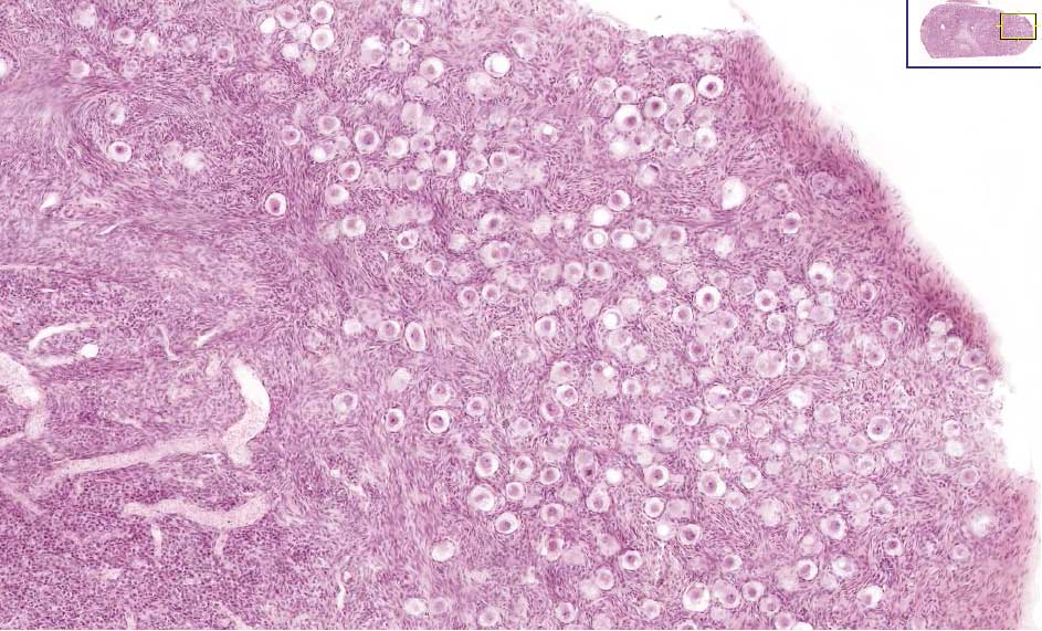
Infant Ovary (human)
In this infant ovary there are a large number of primordial follicles (oocytes) forming a thick cortical region and no later stages follicle development. Compare this structure with the other images of a mature ovary with reproductive activity.
- right - ovary surface (tunica alburginea)
- centre - ovary cortex
- left - ovary medulla
File history
Yi efo/eka'e gwa ebo wo le nyangagi wuncin ye kamina wunga tinya nan
| Gwalagizhi | Nyangagi | Dimensions | User | Comment | |
|---|---|---|---|---|---|
| current | 23:12, 21 September 2009 | | 943 × 571 (108 KB) | S8600021 (talk | contribs) | Infant Ovary, note the large number of primordial follicles around the cortex. |
You cannot overwrite this file.
File usage
The following 24 pages use this file:
- 2009 Lecture 16
- 2010 BGD Practical 3 - Oogenesis and Ovulation
- 2010 Lecture 16
- 2011 Lab 1 - Oogenesis
- 2011 Lab 8 - Postnatal
- 2011 Lecture 16
- 2014 Group Project 4
- ANAT2341 Lab 1 - Oogenesis
- ANAT2341 Lab 8 - Postnatal
- BGDA Lecture - Development of the Embryo/Fetus 1
- BGDA Practical 3 - Oogenesis and Ovulation
- BGDB Sexual Differentiation - Postnatal
- BGD Lecture - Endocrine Development
- BGD Lecture - Sexual Differentiation
- Endocrine - Gonad Development
- In Vitro Oogenesis
- Lecture - Endocrine Development
- Lecture - Fertilization
- Lecture - Genital Development
- Oocyte Development
- Ovary Development
- REI - Reproductive Medicine Seminar 2018
- Royal Hospital for Women - Reproductive Medicine Seminar 2018
- Talk:2014 Group Project 4