File:Fawcett1906 03.jpg
From Embryology
Size of this preview: 605 × 600 pixels. Other resolution: 706 × 700 pixels.
Original file (706 × 700 pixels, file size: 50 KB, MIME type: image/jpeg)

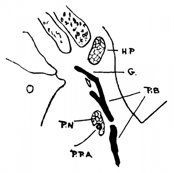
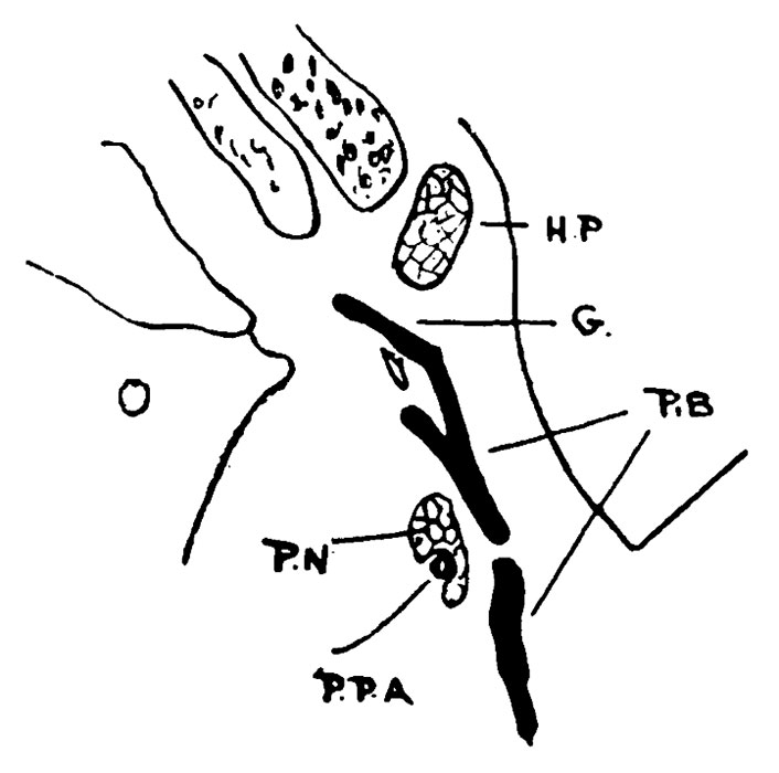
Fig. 3. Palate Embryo
P.B., palate bone; G.,groove for internal pterygoid plate; H.P., hamular process; P.N., palatine nerves; P.P.A., post-palatine artery.
The bone seems here to consist of two separate parts due to foramen.
| Historic Disclaimer - information about historic embryology pages |
|---|
 |
File history
Yi efo/eka'e gwa ebo wo le nyangagi wuncin ye kamina wunga tinya nan
| Gwalagizhi | Nyangagi | Dimensions | User | Comment | |
|---|---|---|---|---|---|
| current | 22:57, 3 April 2014 | | 706 × 700 (50 KB) | Z8600021 (talk | contribs) | ==Fig. 3. Palate Embryo== P.B., palate bone; G.,groove for internal pterygoid plate; H.P., hamular process; P.N., palatine nerves; P.P.A., post-palatine artery. The bone seems here to consist of two separate parts due to foramen. |
You cannot overwrite this file.
File usage
The following page uses this file: