Blood Cells
| Two Blood Cell Systems
|
| Alimentary canal, respiratory passage and urogenital tract.
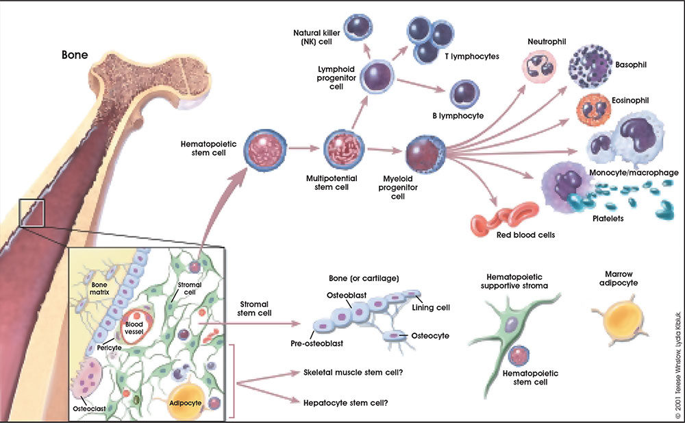
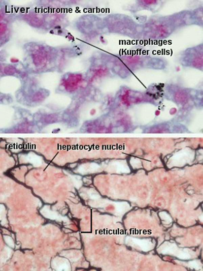
- Mononuclear Phagocytic System - circulating monocytes of peripheral blood and non-circulating (fixed) tissue macrophages found throughout the body.
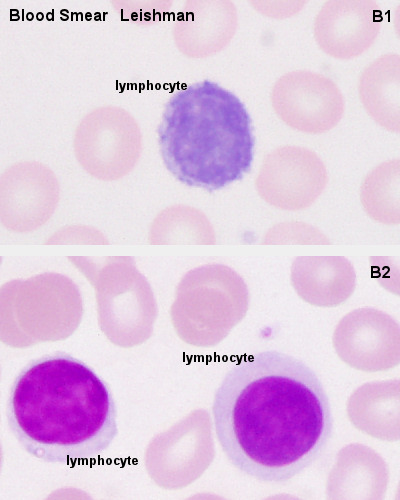
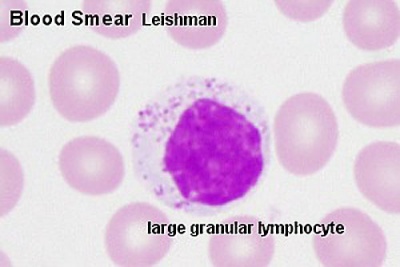
- Lymphoid System - lymphocytes, three major types of T, B, and NK.
Lymphoid Organs
- Central - Lymphocytes develop from precursor cells in bone marrow. (see blood marrow image)
- Peripheral - Lymphocytes respond to antigen lymph nodes or spleen.
| Blood Cells
|
The blood cell information shown below in the table is shown to identify the relative proportions of different cell types in the circulating blood. This information is provided in the lecture as additional information for reference purposes only.
Blood Cell Numbers
The adult ranges of cells / 1 litre (l), total blood volume is about 4.7 to 5 litres. Blood Development | Blood Histology
Red Blood Cells
- Male: 4.32 - 5.66 x 10¹²/l
- Female: 3.88 - 4.99 x 10¹²/l
Leukocytes (white blood cells)
- Male: 3.7 - 9.5 x 10⁹/l
- Female: 3.9 - 11.1 x 10⁹/l
Granulocytes
- 1.8 - 8.9 x 10⁹/l
- Neutrophils: 1.5 - 7.4 x 10⁹/l
- Eosinophils: 0.02 - 0.67 x 10⁹/l
- Basophils: 0 - 0.13 x 10⁹/l
Non-Granulocytes
- Monocytes 0.21 - 0.92 x 10⁹/l
Lymphocytes
- 1.1 - 3.5 x 10⁹/l
- B-cells: 0.06 - 0.66 x 10⁹/l
- T-cells: 0.77 - 2.68 x 10⁹/l
- CD4+: 0.53 - 1.76 x 10⁹/l
- CD8+: 0.30 - 1.03 x 10⁹/l
- NK cells: 0.20 - 0.40 x 10⁹/l
Platelets
- 140 - 440 x 109/l
- not a cell, a cell fragment.
|
|
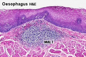
| Mucosal Associated Lymphoid Tissues
|
| Anatomical location - Palatine (tonsils), Lingual and Pharyngeal ( adenoids )
Ring of oral adenoid tissue:
- anterior - lingual tonsil formed by the submucous adenoid collections.
- lateral - palatine tonsils and adenoid collections near the auditory tubes.
- posterior - pharyngeal tonsil on the posterior wall of the pharynx.
- between main masses are smaller collections of adenoid tissue.
|
|
| Tonsils
|
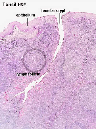
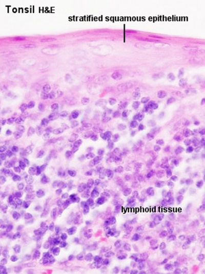
Palatine Tonsils
- the "tonsils", lateral wall of oropharynx
- covered by stratified squamous epithelium
- numerous crypts (10-20) infolds of surface epithelium
- Afferent lymph vessels absent
- Efferent lymph vessels are present
|
|
Lingual Tonsils
- lamina propria root of tongue
- covered by stratified squamous epithelium
- salivary glands and skeletal muscle are directly adjacent
|
Pharyngeal Tonsils
- adenoids or nasopharyngeal tonsils, upper posterior part of throat
- covered by a pseudostratified ciliated epithelium with goblet cells
|
| Peyer's Patch
|
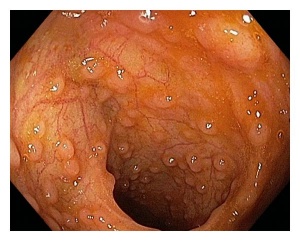
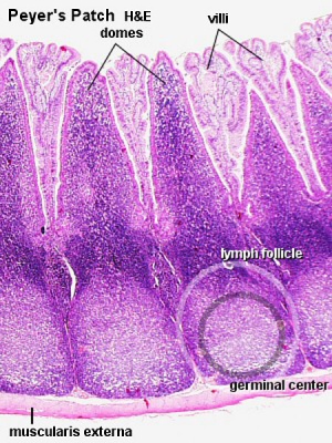
Located in the ileum
|
|
|
| Peyers patches (ileocolonoscopy)
|
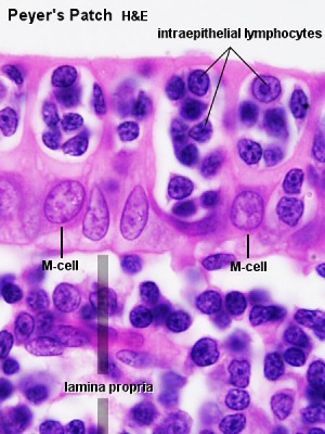
Peyer's Patch (histology)
|
microfold cells or M-cells
(transport gut lumen organisms and particles to immune cells across the epithelial barrier).
|
| About Peyer's Patch
|
| External Links Notice - The dynamic nature of the internet may mean that some of these listed links may no longer function. If the link no longer works search the web with the link text or name. Links to any external commercial sites are provided for information purposes only and should never be considered an endorsement. UNSW Embryology is provided as an educational resource with no clinical information or commercial affiliation.
|
|
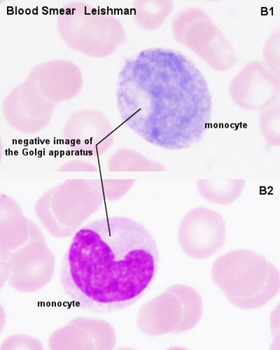
1. Mononuclear Phagocytic System
Mononuclear Phagocytic System (MPS, also called Lymphoreticular System or Reticuloendothelial System, RES)
|
|
Circulating monocytes of peripheral blood.
- monocytes entering the connective tissue differentiate into macrophages)
|
Non-circulating (fixed) tissue macrophages (MΦ)
|
2. Lymphoid System
Adaptive immunity functional cells are the lymphocytes (B, T, NK) and dendritic cells (process antigen and present it on their surface, monocyte precursor derived).
- Antibody-mediated - B Lymphocyte secreting antibody = Plasma Cell
- Cell-mediated - T Lymphocytes form memory cell, Cytotoxic T cells, T helper cell
| B Cell Development
|
Germinal Centres
|
- Bone marrow
- blood
- Lymph node, nodule
- Lymphatic vessel
- Bone marrow
|
- Bone Marrow
- Medullary cords contain plasma cells
|
|
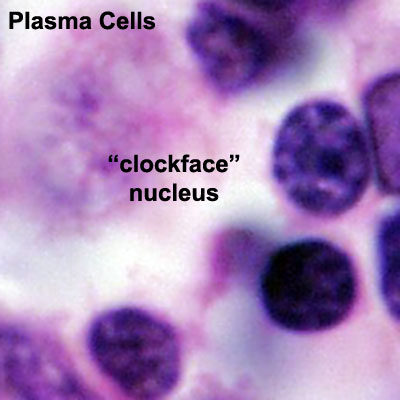
Plasma cells
- Activated B cell, plasma B cells, plasmocytes, effector B cells and B cell that is secreting antibody.
- secrete antibody directly into blood for distribution to all body
- in local extrafollicular sites are short lived 2–4 days
- longer-lived plasma cells in bone marrow 3 weeks to 3 months+
- "clockface" nucleus
- Nucleus has darker (heterochromatin) regions around periphery of nucleus separated by lighter (euchromatin) regions.
|
| Lymphocyte Electron Micrographs
|
| Histologically, there is little difference in appearance between T and B lymphocytes until activated.
|
Lymphocyte Circulation
- Microbial antigens are carried into a lymph node by dendritic cells, which enter via afferent lymphatic vessels draining an infected tissue.
- T and B cells enter the lymph node via an artery and migrate out of the bloodstream through postcapillary venules.
- Unless they encounter their antigen, the T and B cells leave the lymph node via efferent lymphatic vessels, which eventually join the thoracic duct.
- The thoracic duct empties into a large vein carrying blood to the heart.
- A typical circulation cycle takes about 12–24 hours.
Links: MBoC Chapter 24 - The Adaptive Immune System | MBoC Figure 24-14. The path followed by lymphocytes as they continuously circulate between the lymph and blood | Immunobiology


