File:Cooper1932-fig01.jpg
From Embryology
Size of this preview: 435 × 600 pixels. Other resolution: 822 × 1,133 pixels.
Original file (822 × 1,133 pixels, file size: 230 KB, MIME type: image/jpeg)
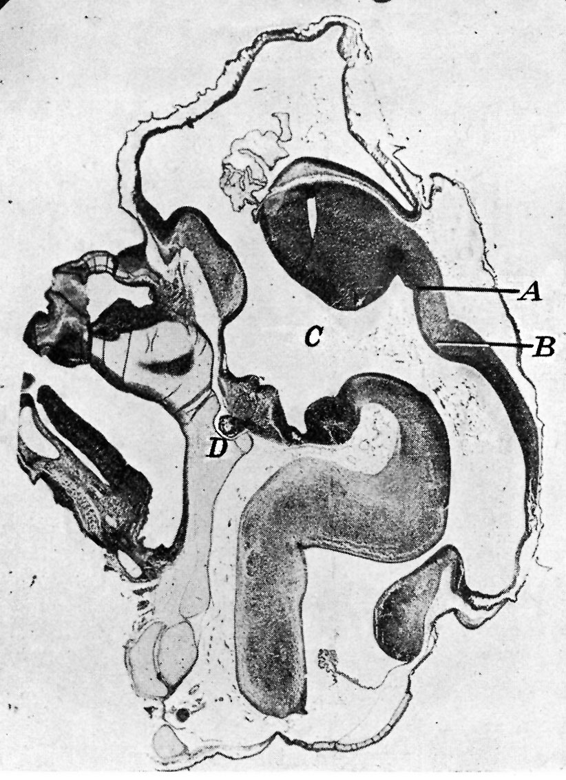
Fig. 1. Sagittal midline section through head of 25 mm embryo
x 8. A, anlage of pineal gland in the form of a backward hollowextension. B, anlage of posterior commissure. C, third ventricle. D, pituitary body.
File history
Yi efo/eka'e gwa ebo wo le nyangagi wuncin ye kamina wunga tinya nan
| Gwalagizhi | Nyangagi | Dimensions | User | Comment | |
|---|---|---|---|---|---|
| current | 18:20, 21 Mayıs 2018 | | 822 × 1,133 (230 KB) | Z8600021 (talk | contribs) | |
| 17:20, 15 November 2015 | 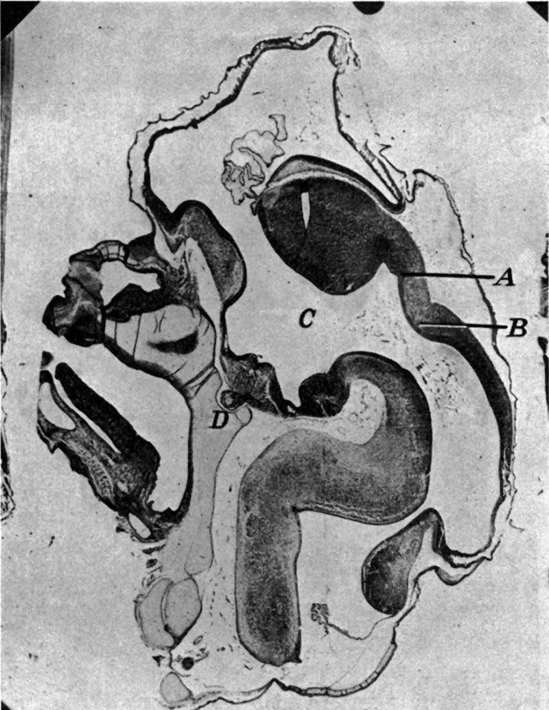 | 800 × 1,035 (184 KB) | Z8600021 (talk | contribs) | ||
| 17:19, 15 November 2015 | 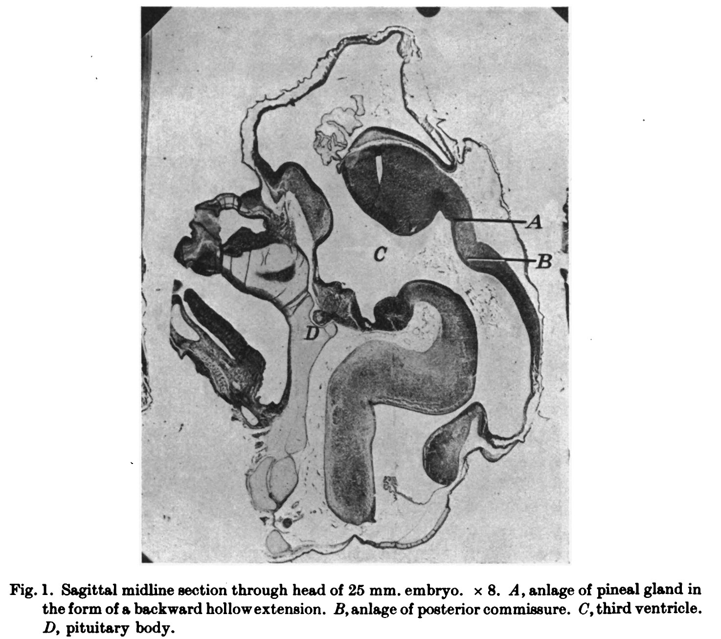 | 1,474 × 1,307 (258 KB) | Z8600021 (talk | contribs) |
You cannot overwrite this file.
File usage
The following 2 pages use this file:


