File:Meyer1932history3 fig01.jpg
From Embryology
Size of this preview: 495 × 599 pixels. Other resolution: 661 × 800 pixels.
Original file (661 × 800 pixels, file size: 147 KB, MIME type: image/jpeg)
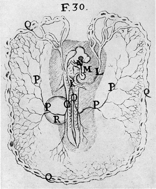
Fig. 1. After Malpighi. Chick after two days’ incubation
K. auricle; L. veins; M, right ventricle; N, left sinus; O, branches of umbilical arteries; P, umbilical arteries; Q, terminations of arteries; R, vesicle near right umbilical branch P is swollen with thin humor or ichor. (This vesicle must have been the extremity of the allantois.)
File history
Yi efo/eka'e gwa ebo wo le nyangagi wuncin ye kamina wunga tinya nan
| Gwalagizhi | Nyangagi | Dimensions | User | Comment | |
|---|---|---|---|---|---|
| current | 18:07, 2 November 2015 | | 661 × 800 (147 KB) | Z8600021 (talk | contribs) | |
| 18:07, 2 November 2015 | 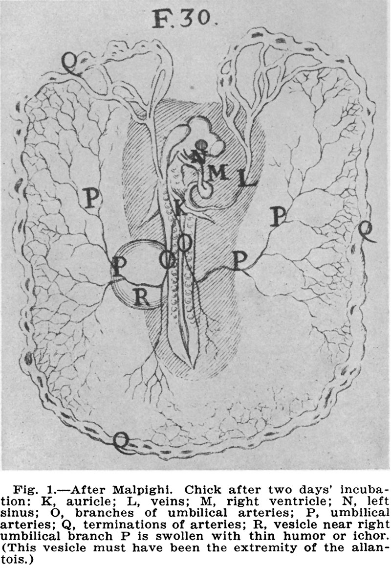 | 800 × 1,163 (215 KB) | Z8600021 (talk | contribs) |
You cannot overwrite this file.
File usage
The following page uses this file:


