File:Bardeen1908 fig1B.jpg
From Embryology
Size of this preview: 488 × 600 pixels. Other resolution: 1,496 × 1,838 pixels.
Original file (1,496 × 1,838 pixels, file size: 280 KB, MIME type: image/jpeg)
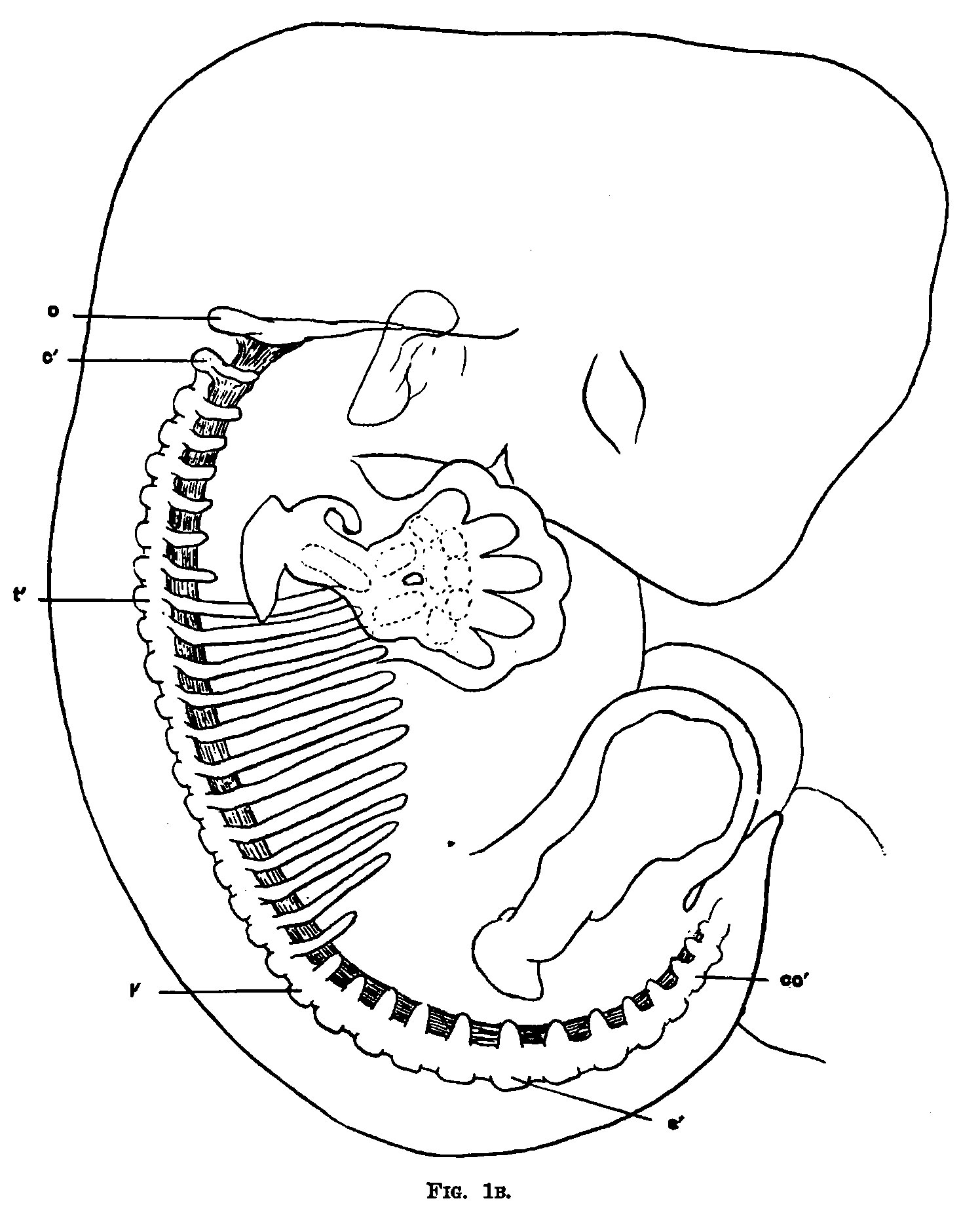
Fig. 1B. Diagram of the skeleton of an embryo 11 mm long and about five weeks old
0, occipital plate. 1'. first lumbar vertebra. c, flrst cervical vertebra. s', first sacral vertebra. t’, that thoracic vertebra. co’, first coccygeal vertebra.
Cite this page: Hill, M.A. (2026, October 5) Embryology Bardeen1908 fig1B.jpg. Retrieved from https://embryology.med.unsw.edu.au/embryology/index.php/File:Bardeen1908_fig1B.jpg
- © Dr Mark Hill 2026, UNSW Embryology ISBN: 978 0 7334 2609 4 - UNSW CRICOS Provider Code No. 00098G
File history
Yi efo/eka'e gwa ebo wo le nyangagi wuncin ye kamina wunga tinya nan
| Gwalagizhi | Nyangagi | Dimensions | User | Comment | |
|---|---|---|---|---|---|
| current | 18:48, 28 October 2015 | | 1,496 × 1,838 (280 KB) | Z8600021 (talk | contribs) | |
| 18:48, 28 October 2015 | 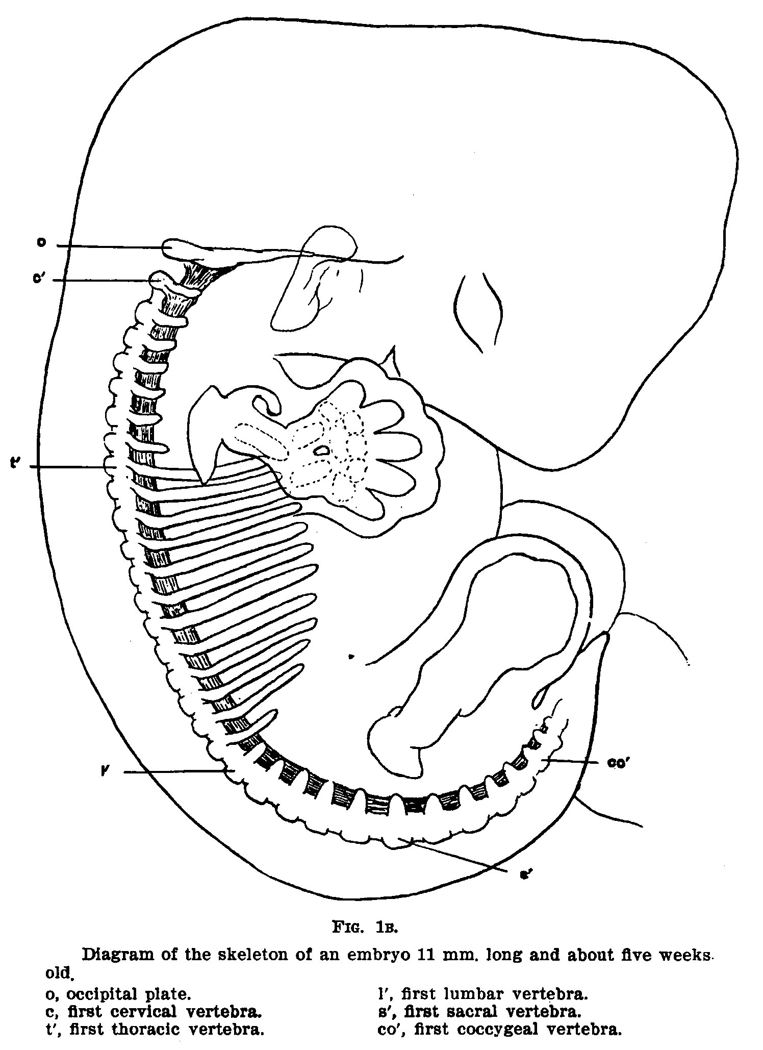 | 1,496 × 2,058 (341 KB) | Z8600021 (talk | contribs) |
You cannot overwrite this file.
File usage
The following page uses this file:


