File:Vaginal Ultrasonography in Sagittal Plane.jpg
From Embryology
Size of this preview: 800 × 455 pixels. Other resolution: 975 × 554 pixels.
Original file (975 × 554 pixels, file size: 112 KB, MIME type: image/jpeg)
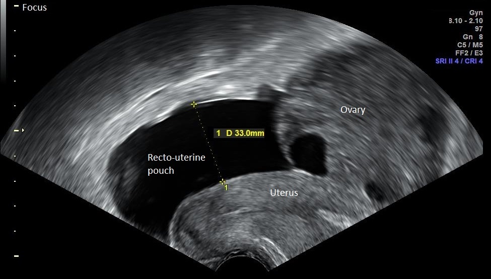
Image showing the vaginal ultrasonography, sagittal section, of a woman who has been diagnosed with mild ovarian hyperstimulation syndrome. Fluid from the ovaries has filled the recto-uterine pouch.
File history
Yi efo/eka'e gwa ebo wo le nyangagi wuncin ye kamina wunga tinya nan
| Gwalagizhi | Nyangagi | Dimensions | User | Comment | |
|---|---|---|---|---|---|
| current | 00:04, 21 October 2015 | | 975 × 554 (112 KB) | Z3374116 (talk | contribs) | Image showing the vaginal ultrasonography, sagittal section, of a woman who has been diagnosed with mild ovarian hyperstimulation syndrome. Fluid from the ovaries has filled the recto-uterine pouch. |
You cannot overwrite this file.
File usage
The following 2 pages use this file:


