File:Hamilton1945-plate01.jpg
Original file (1,490 × 2,000 pixels, file size: 478 KB, MIME type: image/jpeg)
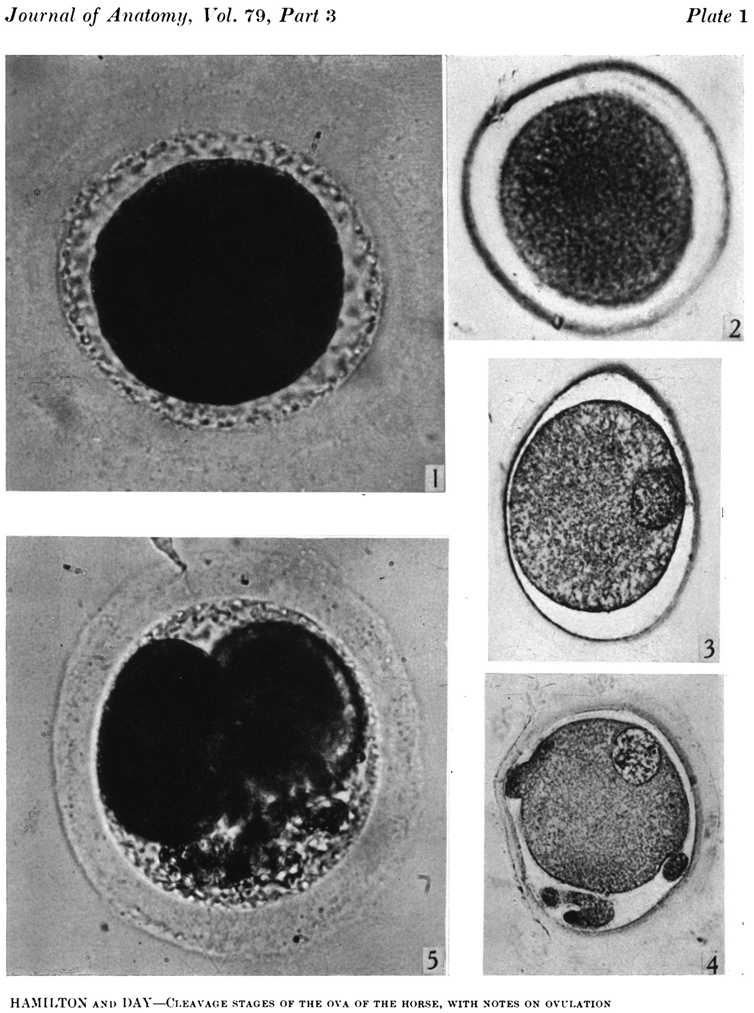
Plate 1
Fig. 1. Photograph of a living unsegmented ovum of the pony (P 1) in Locke’s fluid. Granular material is seen in the perivitelline space. X 480.
Fig. 2: Photograph of an unsegmented ovum of the pony (P 1) in agar. The nucleus is just visible. x 480.
Fig. 3. Section of an unsegmented ovum of the pony (P1) with a large vesicular nucleus. x 520.
Fig. 4. Section of an unsegmented ovum of the pony (P 8) showing deutoplasmolysis. x 520.
Fig. 5. Photograph of a living two-cell stage ovum of the pony (P 5). Granular material is seen in the lower part of the perivitelline space. x 480.
| Historic Disclaimer - information about historic embryology pages |
|---|
 |
- Links: Fig. 1 | Fig. 2 | Fig. 3 | Fig. 4 | Fig. 5 | Plate 1 | Fig 6 | Fig 7 | Fig 8 | Fig 9 | Fig 10 | Fig 11 | Plate 2 | Fig 12 | Fig 13 | Fig 14 | Fig 15 | Plate 3 | Hamilton 1945 | Historic Embryology Papers | Oocyte Development | Morula | Horse Development | Animal Development
Reference
Hamilton WJ. Cleavage stages of the ova of the horse, with notes on ovulation. (1945) J Anat. 79(3): 127–130. PMID 17104976
Cite this page: Hill, M.A. (2024, May 23) Embryology Hamilton1945-plate01.jpg. Retrieved from https://embryology.med.unsw.edu.au/embryology/index.php/File:Hamilton1945-plate01.jpg
- © Dr Mark Hill 2024, UNSW Embryology ISBN: 978 0 7334 2609 4 - UNSW CRICOS Provider Code No. 00098G
File history
Click on a date/time to view the file as it appeared at that time.
| Date/Time | Thumbnail | Dimensions | User | Comment | |
|---|---|---|---|---|---|
| current | 11:36, 29 September 2015 | | 1,490 × 2,000 (478 KB) | Z8600021 (talk | contribs) | ===Plate 1=== Fig. 1. Photograph of a living unsegmented ovum of the pony (P 1) in Locke’s fluid. Granular material is seen in the perivitelline space. X 480. Fig. 2: Photograph of an unsegmented ovum of the pony (P 1) in agar. The nucleus is just... |
You cannot overwrite this file.
File usage
The following 3 pages use this file: