File:Hamilton1945-plate01.jpg
Original file (1,490 × 2,000 pixels, file size: 478 KB, MIME type: image/jpeg)
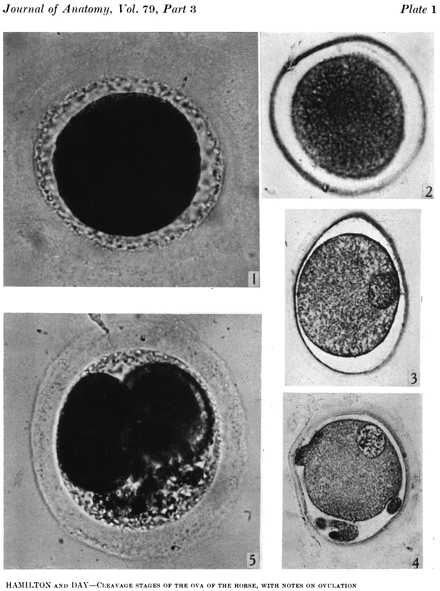
Plate 1
Fig. 1. Photograph of a living unsegmented ovum of the pony (P 1) in Locke’s fluid. Granular material is seen in the perivitelline space. X 480.
Fig. 2: Photograph of an unsegmented ovum of the pony (P 1) in agar. The nucleus is just visible. x 480.
Fig. 3. Section of an unsegmented ovum of the pony (P1) with a large vesicular nucleus. x 520.
Fig. 4. Section of an unsegmented ovum of the pony (P 8) showing deutoplasmolysis. x 520.
Fig. 5. Photograph of a living two-cell stage ovum of the pony (P 5). Granular material is seen in the lower part of the perivitelline space. x 480.
File history
Yi efo/eka'e gwa ebo wo le nyangagi wuncin ye kamina wunga tinya nan
| Gwalagizhi | Nyangagi | Dimensions | User | Comment | |
|---|---|---|---|---|---|
| current | 11:36, 29 September 2015 | | 1,490 × 2,000 (478 KB) | Z8600021 (talk | contribs) | ===Plate 1=== Fig. 1. Photograph of a living unsegmented ovum of the pony (P 1) in Locke’s fluid. Granular material is seen in the perivitelline space. X 480. Fig. 2: Photograph of an unsegmented ovum of the pony (P 1) in agar. The nucleus is just... |
You cannot overwrite this file.
File usage
The following 3 pages use this file: