File:Streeter1917-fig04-05.jpg
Original file (1,000 × 544 pixels, file size: 124 KB, MIME type: image/jpeg)
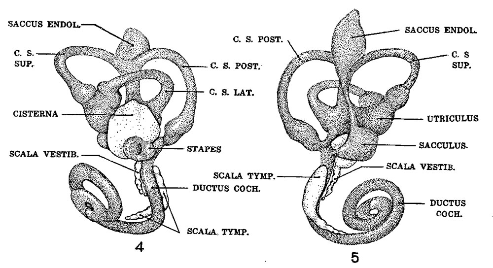
Fig. 4 The figures 4 to 9 represent a series of median and lateral views of wax-plate reconstructions of the membranous labyrinth and the surrounding perioticular tissue spaces, illustrating under the same scale of enlargement three typical stages in the development of these spaces. This figure shows a lateral view of a model reconstructed from a human fetus 50 mm. CR length (Carnegie Collection, No. 84). The scala vestibuli is in the first stage of its development and consists of a row of large reticular spaces which extend from the ventral margin of the cistern downward along the apical surface of the cochlear duct. The scala tympani is more advanced and shows more complete coalescence of its constituent spaces. Enlarged 9 diameters.
Fig. 5 Median View of the same model shown in figure 4. This view shows the topography of the scala tympani. Its large proximal end lies opposite the
fenestra cochleac and corresponds to the focus at which its development originates. Distally it tapers off rapidly, where the spaces are smaller and their
coalescence less complete. Enlarged 9 diameters.
File history
Yi efo/eka'e gwa ebo wo le nyangagi wuncin ye kamina wunga tinya nan
| Gwalagizhi | Nyangagi | Dimensions | User | Comment | |
|---|---|---|---|---|---|
| current | 16:04, 17 September 2015 | | 1,000 × 544 (124 KB) | Z8600021 (talk | contribs) | |
| 16:03, 17 September 2015 |  | 1,350 × 1,329 (472 KB) | Z8600021 (talk | contribs) |
You cannot overwrite this file.
File usage
There are no pages that use this file.


