File:Ovary histology with chemotherapy.jpg
From Embryology
Size of this preview: 672 × 600 pixels. Other resolution: 977 × 872 pixels.
Original file (977 × 872 pixels, file size: 232 KB, MIME type: image/jpeg)
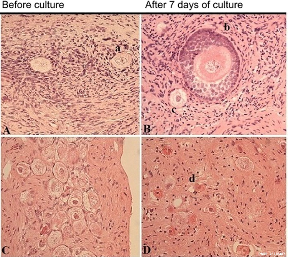
Representative images of ovarian cortex before and after seven days of culture (magnification ×40), from a 15-year-old girl with lymphoma and no chemotherapy (A,B), and from a 2-year-old girl with neuroblastoma exposed to CED of 7200 mg/m2 (C,D). a) Intact primordial follicle, b) intact secondary follicle, c) influenced primordial follicle, d) atretic follicle.
Journal.pone.0133985.g001.jpg
File history
Yi efo/eka'e gwa ebo wo le nyangagi wuncin ye kamina wunga tinya nan
| Gwalagizhi | Nyangagi | Dimensions | User | Comment | |
|---|---|---|---|---|---|
| current | 11:40, 5 September 2015 | | 977 × 872 (232 KB) | Z8600021 (talk | contribs) | Representative images of ovarian cortex before and after seven days of culture (magnification ×40), from a 15-year-old girl with lymphoma and no chemotherapy (A,B), and from a 2-year-old girl with neuroblastoma exposed to CED of 7200 mg/m2 (C,D). a) I... |
You cannot overwrite this file.
File usage
The following page uses this file:


