File:Thompson1908 02.jpg
From Embryology

Size of this preview: 800 × 417 pixels. Other resolution: 1,444 × 753 pixels.
Original file (1,444 × 753 pixels, file size: 192 KB, MIME type: image/jpeg)
Fig. 2. Schematic representation of three sections of embryo
A, at level of sinus venosus:
B, at level of liver bud;
C, at level of ventral mesentery.
N.,notochord ; Ao.,aoI-ta; S.V.,sinus venosus; S.'l‘.,septum transversum ; C.,ca-rlom; D.C., ductus Cuv.; V., ventricle; 1>.C., pericanlial cavity; 8., stomach; H.B., liver bud; V.V., vltelline vein; U.V., umbilical vein; Am., amnion; V.M., ventral mesentery; 1., small intestine.
File history
Yi efo/eka'e gwa ebo wo le nyangagi wuncin ye kamina wunga tinya nan
| Gwalagizhi | Nyangagi | Dimensions | User | Comment | |
|---|---|---|---|---|---|
| current | 23:56, 11 August 2015 | | 1,444 × 753 (192 KB) | Z8600021 (talk | contribs) | |
| 23:56, 11 August 2015 | 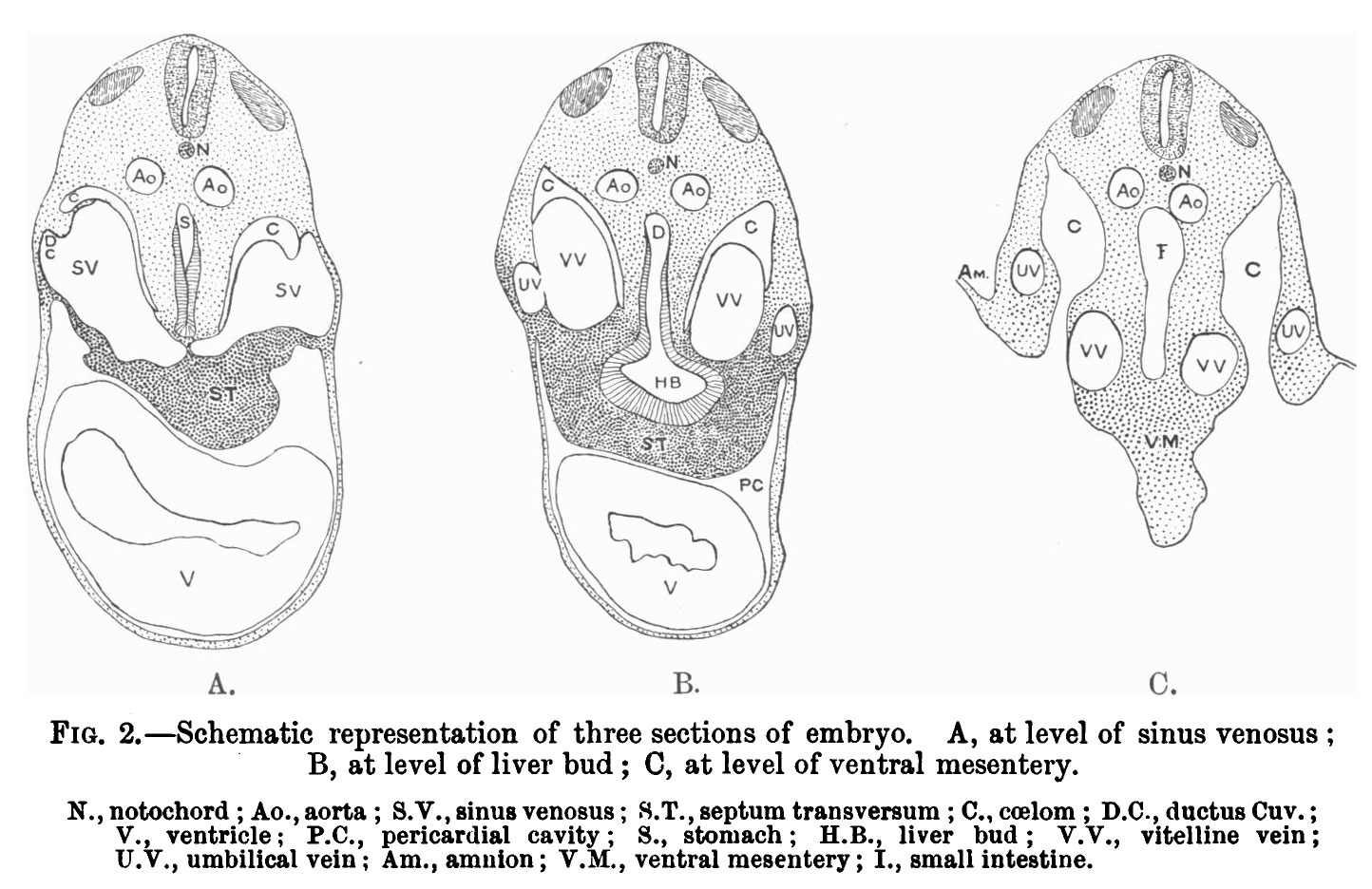 | 1,444 × 936 (236 KB) | Z8600021 (talk | contribs) | ==Fig. 2. Schematic representation of three sections of embryo== A, at level of sinus venosus: B, at level of liver bud; O, at level of ventral mesentery. N.,notochord ; Ao.,aoI-ta; S.V.,sinus venosus; S.'l‘.,septum transversum ; C.,ca-rlom; D.C.,... |
You cannot overwrite this file.
File usage
There are no pages that use this file.


