File:Urinary Bladder Histology.jpg
Urinary_Bladder_Histology.jpg (581 × 399 pixels, file size: 42 KB, MIME type: image/jpeg)
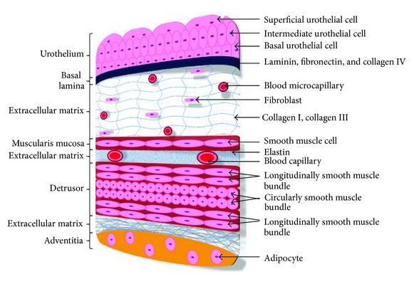
The histology of the urinary bladder showing the different cell layers
<pubmed>24453796</pubmed>
Copyright © 2013 Hazem Orabi et al. This is an open access article distributed under the Creative Commons Attribution License, which permits unrestricted use, distribution, and reproduction in any medium, provided the original work is properly cited.
- Note - This image was originally uploaded as part of an undergraduate science student project and may contain inaccuracies in either description or acknowledgements. Students have been advised in writing concerning the reuse of content and may accidentally have misunderstood the original terms of use. If image reuse on this non-commercial educational site infringes your existing copyright, please contact the site editor for immediate removal.
--Mark Hill (talk) 12:34, 11 October 2014 (EST) Please avoid using all capitals in file names and should also be called "cartoon" as it is not an actual histology image. Please also add sub-headings for reference and copyright above. I have also added additional categories to your uploaded image, it is not part of the final assessment.
File history
Yi efo/eka'e gwa ebo wo le nyangagi wuncin ye kamina wunga tinya nan
| Gwalagizhi | Nyangagi | Dimensions | User | Comment | |
|---|---|---|---|---|---|
| current | 22:39, 8 October 2014 | | 581 × 399 (42 KB) | Z5030311 (talk | contribs) | The histology of the urinary bladder showing the different cell layers <pubmed>24453796</pubmed> Copyright © 2013 Hazem Orabi et al. This is an open access article distributed under the Creative Commons Attribution License, which permits unrestricte... |
You cannot overwrite this file.
File usage
The following 2 pages use this file:


