File:Limbal palisades of Vogt PMID17211449.jpg
Original file (692 × 800 pixels, file size: 86 KB, MIME type: image/jpeg)
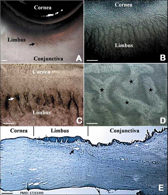
The limbal palisades of Vogt
Palisades of Vogt (arrow) are readily recognized in the human limbus (A). Such a unique pigmented structure can be identified on the flat mount preparation of Dispase-isolated human limbal epithelial sheets (B). In donors with a darker skin, these palisades of Vogt are pigmented (C, arrow). Under higher magnification, these limbal areas show undulated epithelial papillae (D, stars). Hematoxyline staining highlights higher stratification and more undulation of the limbal epithelium, and the underlying limbal stroma has high cellularity and vascularity (E, arrow shows blood vessel). (Bar represents 500 μm in A and B, 200 μm in C and E, and 50 μm in D)
File history
Yi efo/eka'e gwa ebo wo le nyangagi wuncin ye kamina wunga tinya nan
| Gwalagizhi | Nyangagi | Dimensions | User | Comment | |
|---|---|---|---|---|---|
| current | 12:11, 30 August 2014 | | 692 × 800 (86 KB) | Z8600021 (talk | contribs) | ==The limbal palisades of Vogt== Palisades of Vogt (arrow) are readily recognized in the human limbus (A). Such a unique pigmented structure can be identified on the flat mount preparation of Dispase-isolated human limbal epithelial sheets (B). In don... |
You cannot overwrite this file.
File usage
The following page uses this file: